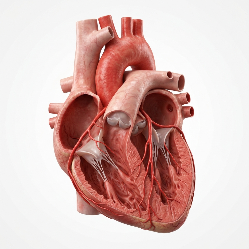
Aortic Stenosis
Aortic valve stenosis is the progressive narrowing of the aortic valve orifice. Modern transcatheter valve replacement (TAVR) has revolutionised treatment, offering exceptional recovery and outcomes.
The Pathology of Aortic Valve Narrowing
Aortic stenosis restricts the flow of blood out of the heart's primary pumping chamber, leading to severe pressure loads on the ventricle.
The normal aortic valve opening is between 3.0 to 4.0 cm². Degenerative calcification narrows this space. When it reaches severe parameters (<1.0 cm²), the heart can no longer compensate, triggering rapid symptom onset.
Causes & Risk Factors for Aortic Stenosis
Aortic stenosis arises from degenerative, congenital, or rheumatic processes.
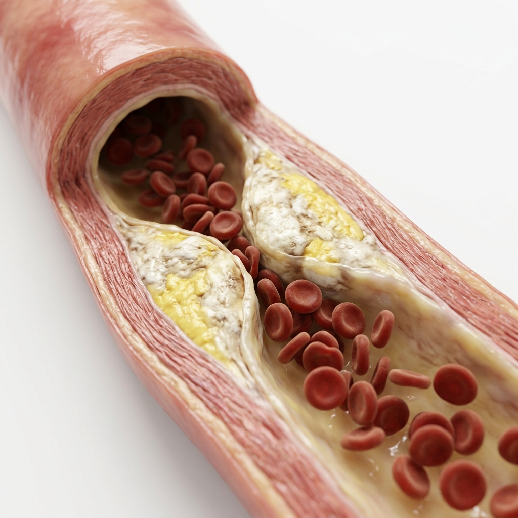
Degenerative Calcific AS
Age-related calcification of a trileaflet valve — most common cause in patients >70 years. Risk factors include hypertension, diabetes, hyperlipidaemia, and smoking.
Bicuspid Aortic Valve (BAV)
Congenital condition where the valve has two leaflets instead of three. Present in 1–2% of the population. Causes AS 2–3 decades earlier (age 50–65). Associated with ascending aortopathy.
Rheumatic Heart Disease
Rheumatic valve damage following childhood streptococcal infection. Causes combined AS and AR (aortic regurgitation). More common in developing countries.
Chronic Kidney Disease
CKD accelerates vascular and valvular calcification due to disordered calcium-phosphate metabolism. AS progresses faster in dialysis patients.
Genetic Factors
NOTCH1 gene mutation associated with bicuspid valve and early calcification. Lp(a) elevation is an independent risk factor for AS progression.
Radiation Therapy
Previous mediastinal radiation (for lymphoma, breast cancer) causes accelerated valve calcification and fibrosis, typically 10–20 years after treatment.
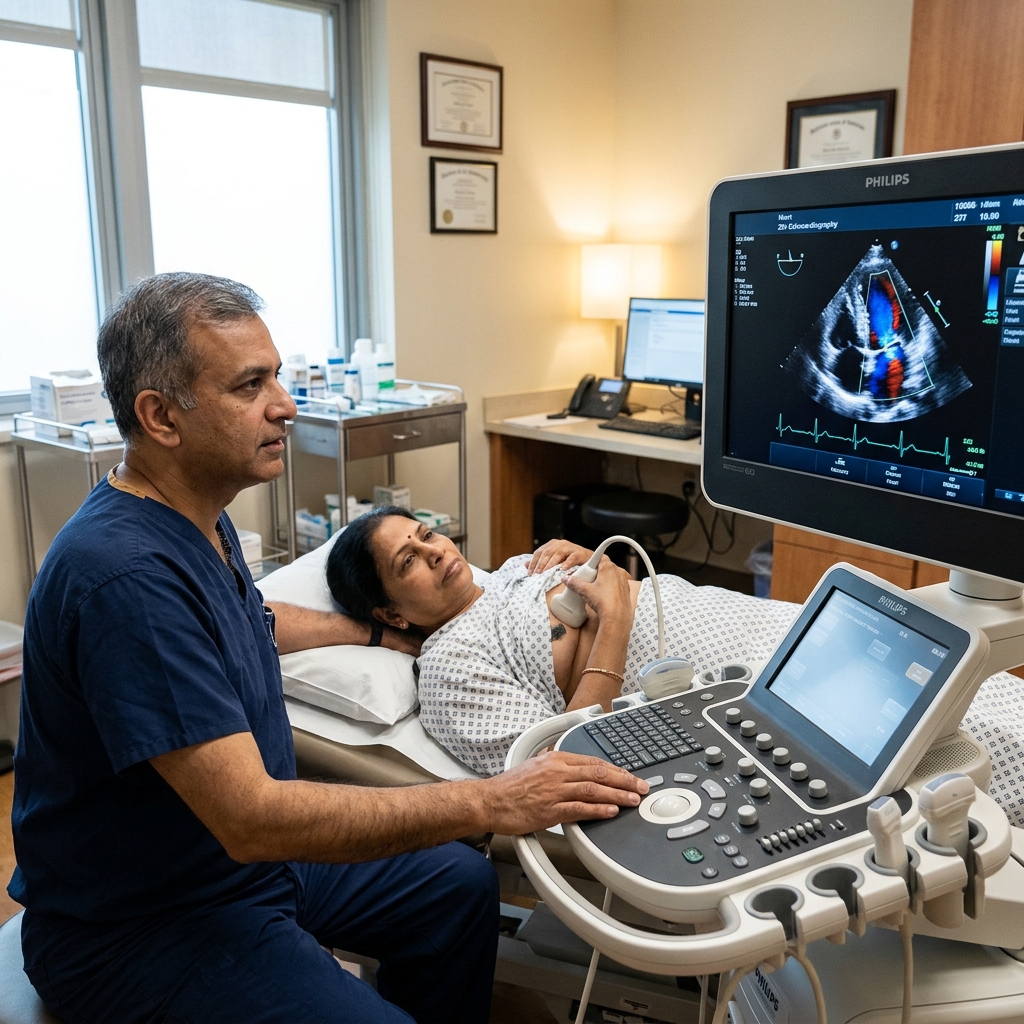
Echo Grading Guidelines
We grade aortic stenosis severity precisely via multi-parameter Doppler echocardiography parameters.
The Symptom Triad: Survival Timelines
Aortic stenosis progresses silently, but once physical symptoms present, survival drops dramatically without mechanical repair.
Angina
~3 years survivalMedian survival after onset of exertional chest pain is ~3 years. 50% of patients die within 3 years without intervention. Chest pain results from LV pressure overload and increased oxygen demand.
Syncope
~2–3 years survivalExertional fainting occurs when cardiac output cannot increase adequately. Sudden cardiac death risk is high. Median survival is ~2–3 years after onset.
Breathlessness (HF)
~1–2 years survivalWorst prognosis — indicates the LV has decompensated. Survival is 50% at 1–2 years without urgent valve replacement.
How Is Aortic Stenosis Diagnosed?
Doppler echocardiography is the definitive diagnostic tool for aortic stenosis.
Transthoracic Echocardiography (TTE)
Measures aortic valve area (AVA) by continuity equation, peak velocity across the valve (Vmax), and mean gradient. TTE is the primary diagnostic and surveillance tool.
CT Aortogram & Valve Scoring
Quantifies valve calcification (Agatston score — >2000 AU in men, >1200 AU in women indicates severe AS). Assesses annular dimensions for TAVR planning and aortopathy.
Exercise Testing
In asymptomatic severe AS, treadmill test unmask symptoms or abnormal BP response. A positive test triggers earlier valve intervention (Class IIa).
Cardiac Catheterisation
Direct measurement of transvalvular gradient. Rarely needed — reserved when non-invasive tests are discordant or inconclusive.
BNP / NT-proBNP
Elevated BNP in AS correlates with symptom severity and LV decompensation. Serial measurement helps time intervention in asymptomatic patients.
Natural History Without Intervention
Aortic stenosis follows a predictable downhill course once symptoms develop.
Angina — Onset to Death ~3 Years
Exertional chest pain marks the beginning of the symptomatic phase. Without valve replacement, median survival is approximately 3 years.
Syncope — Onset to Death ~2–3 Years
Fainting on exertion indicates fixed cardiac output. Risk of sudden cardiac death is elevated.
Heart Failure — Onset to Death ~1–2 Years
Breathlessness is the most ominous symptom. It means the left ventricle has decompensated and cannot maintain adequate output.
Sudden Cardiac Death
AS carries a risk of sudden death, especially during the symptomatic phase. This is prevented by timely valve replacement.
ESC & AHA Intervention Criteria
Clear, evidence-backed clinical triggers that determine when a patient must undergo valve replacement.
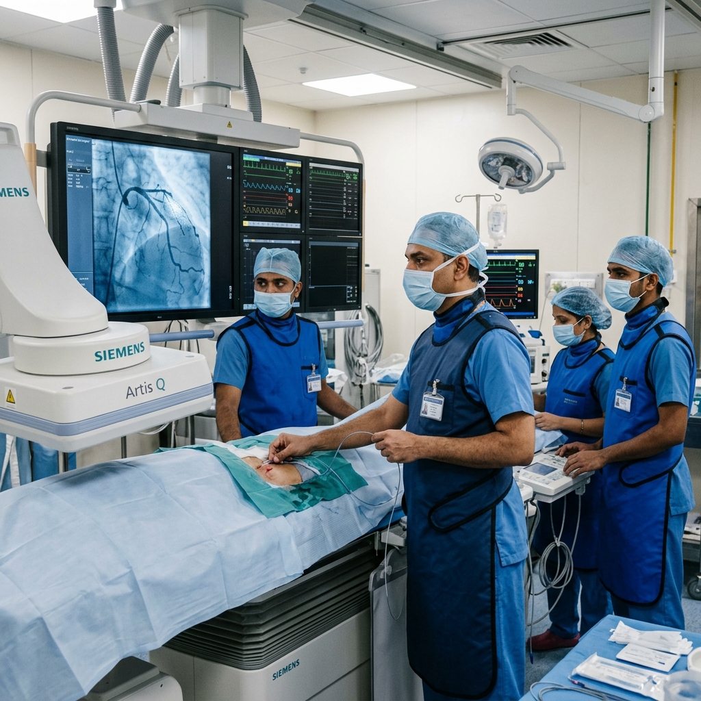
TAVR (Transcatheter AVR)
PARTNER 3: 1.0% vs 3.3% 30-day compositeCatheter-based valve delivery via femoral artery. No chest incision, shorter recovery. PARTNER 3 proved TAVR superiority in low-risk patients.
SAVR (Surgical AVR)
Durable surgical valvesOpen-chest valve replacement. Preferred in young patients (<65), bicuspid valve, and when concomitant CABG or other cardiac surgery is needed.
Class I — Mandatory
ESC/AHA Class ISevere AS + any symptoms (angina, syncope, breathlessness). Severe AS + LVEF <50%. Very severe AS + high gradient. Valve replacement as soon as feasible.
Medications for Aortic Stenosis
No medication has been proven to slow AS progression or improve outcomes in severe disease.
Lifestyle with Aortic Stenosis
Patients with AS should avoid strenuous activity and maintain regular follow-up.
Avoid Strenuous Exertion
Modify activity levelAvoid heavy lifting, competitive sports, and maximal exertion. These cause sudden pressure load on the LV potentially triggering syncope or sudden death.
Regular Echo Surveillance
Essential monitoringEcho every 6–12 months for severe AS, 1–2 years for moderate. Report any new symptoms immediately.
Heart-Healthy Diet
Risk factor controlControl BP, cholesterol, and diabetes. Mediterranean diet recommended. Salt restriction if hypertension present.
Symptom Awareness
Emergency warning signsReport any new chest pain, shortness of breath, or fainting immediately. These are triggers for urgent valve evaluation.
Definitive Clinical Trials
The safety and efficacy of TAVR versus open-chest surgery has been proven across extensive multi-centre randomized trials.
When to See a Doctor
Certain symptoms in AS require immediate medical evaluation.
New Chest Pain (Angina)
Onset of exertional chest pain in a patient with known AS requires urgent evaluation — median survival is only ~3 years without intervention.
Fainting (Syncope)
Exertional syncope indicates advanced AS with fixed cardiac output. Risk of sudden death is high. Emergency evaluation is required.
Worsening Breathlessness
Progressive dyspnoea or orthopnoea signals LV decompensation. Urgent valve replacement is typically indicated.
Rapid Progression on Echo
If annual echo shows a rapid increase in gradient (>20 mmHg/year), intervention may be needed even without symptoms.
Frequently Asked Questions
Detailed, peer-reviewed clarifications regarding aortic valve stenosis and TAVR options.
Aortic stenosis (AS) is the progressive narrowing of the aortic valve — the valve between the left ventricle and the aorta — due to calcification and fibrosis of the valve leaflets. As the valve narrows, the left ventricle must generate progressively higher pressures to force blood through the obstructed opening, leading to hypertrophy, diastolic dysfunction, and eventually failure.
The classic triad of severe aortic stenosis symptoms consists of: 1) Angina — chest pain on exertion; 2) Syncope — fainting on exertion; and 3) Breathlessness (dyspnoea) — the earliest symptom of heart failure. Once symptoms develop, survival without valve intervention deteriorates rapidly (1 to 3 years median survival).
TAVR (Transcatheter Aortic Valve Replacement) is a catheter-based procedure — a new valve is delivered via the femoral artery and expanded within the diseased native valve, without opening the chest. SAVR requires open-chest surgery. PARTNER 3 (NEJM 2019) demonstrated TAVR's superiority or equivalence across all surgical risk groups.
No medication has been proven to slow the progression of aortic stenosis or improve outcomes once the valve is significantly narrowed. Once severe symptomatic AS develops, valve replacement (TAVR or SAVR) is the only effective treatment.
A bicuspid aortic valve is a congenital heart condition where the aortic valve has two leaflets instead of the normal three. It is the most common congenital heart defect, affecting approximately 1–2% of the population. People with bicuspid valves develop aortic stenosis earlier in life — typically in their 50s or 60s, compared to 70s or 80s for patients with trileaflet valves who develop degenerative calcific stenosis. Bicuspid valves are also associated with aortic root dilation and increased risk of aortic dissection. Anyone with a bicuspid valve needs lifelong echocardiographic surveillance of both valve function and ascending aortic size, even if the valve is functioning normally.
Currently, no medication has been proven to prevent or slow the progression of aortic stenosis once the valve starts to calcify — this is different from coronary artery disease, where statins and lifestyle changes are effective. Early studies suggested statins might help, but large trials (SALTIRE, SEAS) did not demonstrate any benefit in slowing stenosis progression. The only definitive treatment remains valve replacement — either surgical (SAVR) or transcatheter (TAVR) — once symptoms develop or the stenosis becomes severe. However, aggressive treatment of coexisting hypertension and coronary artery disease is essential, as these conditions increase the workload on the heart and worsen outcomes in patients with aortic stenosis.
TAVR valves have demonstrated excellent durability. The 5-year and 8-year follow-up data from the PARTNER trials show that TAVR valves maintain good haemodynamic function with no evidence of structural deterioration in the majority of patients. The NOTION trial, which randomised low-risk patients to TAVR vs SAVR, reported that at 10 years, bioprosthetic valve failure rates were similar between TAVR and surgical valves (approximately 10–15%). For patients over 75, a TAVR valve is expected to last the rest of their life in most cases. For younger patients, a TAVR valve may eventually require a second procedure (valve-in-valve TAVR), which is a well-established and safe option.
Exercise recommendations depend on the severity of the stenosis. Patients with mild aortic stenosis and no symptoms can participate in all forms of exercise without restriction, although annual echocardiographic follow-up is recommended. Patients with moderate stenosis should avoid competitive sports and heavy weightlifting but can engage in moderate aerobic exercise such as walking, light cycling, and swimming. Patients with severe aortic stenosis should avoid strenuous exercise and any competitive sports — even if they feel well — as sudden cardiac death during exertion is a known risk. Once the valve is replaced (TAVR or SAVR), most patients can return to normal physical activity, including exercise, after appropriate recovery and cardiac rehabilitation.
“Advanced cardiovascular care. Restoring life, rhythm, and vitality.”

Dr. Amit Singh, FACC
Consultant Interventional Cardiologist
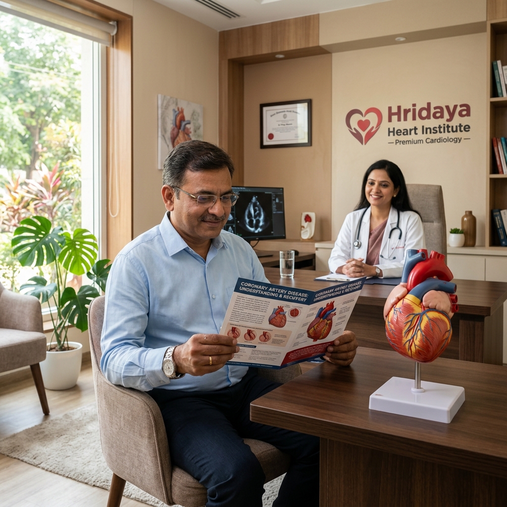
Cardiac Services Available at Heartwise
Dr. Amit Singh offers comprehensive cardiac diagnostics and management for this condition at Heartwise Cardiology Clinic, Vashi.
TAVR Valve Replacement
Minimally invasive transcatheter aortic valve replacement without open surgery.
2D Echocardiography
Detailed Doppler assessment of AVA, mean gradient, and LV function.
Coronary Artery Disease
CAD frequently coexists with AS — pre-TAVR coronary assessment is routine.
Heart Failure
Advanced AS leads to HF; comprehensive post-intervention HF monitoring.
Book a Visit.
Pick a date and time that works for you.
Select a date
| Su | Mo | Tu | We | Th | Fr | Sa |
|---|---|---|---|---|---|---|
Book an Appointment with Dr. Amit Singh, FACC.
Schedule a cardiology consultation at Heartwise Clinic in Vashi, Navi Mumbai — online booking, WhatsApp, or call. Dr. Amit Singh offers in-clinic and secure video teleconsultations for patients across India and internationally.
Choose Date & Time
Pick a slot that fits your schedule from available morning or evening appointments.
Share Your Details
Provide your name, contact number, and a brief note about your cardiac concern or reason for visit.
Get Confirmed
Our clinical team confirms your slot within 24 hours via call or WhatsApp with pre-visit instructions.
Consult In-Clinic or Online
Visit Heartwise Clinic in Vashi or join a secure HD video teleconsultation from anywhere.
Consultation Options
In-Clinic Consultation
Kokilaben Hospital, Kopar Khairane & Heartwise Clinic, Vashi
HD Video Teleconsultation
Available pan-India and for international patients
WhatsApp Booking
Quick booking via +91 97695 17636 — reports & follow-ups
100+ appointments this month
Confirmed by our clinical team
4.9 / 5 rating
From patient reviews
Our Cardiology
Centers.
Dr. Amit Singh consults across multiple flagship centers and outreach clinics in Navi Mumbai & Dombivli to ensure specialized, top-tier cardiac care is directly accessible.
Navi Mumbai Sectors & Surrounding Nodes Served
Triple ESC & FACC Certified
International guidelines and clinical safety protocols applied across all heart centers.
“Beat Better. Live Wiser.”
Dr. Amit Singh, FACC
Consultant Interventional Cardiologist
Medical Disclaimer: This article has been written and reviewed by Dr. Amit Singh, FACC, for educational purposes only. It does not constitute personalised medical advice and should not be used as a substitute for a consultation with a qualified cardiologist. Individual clinical decisions must be made by a treating physician based on complete medical history and examination. If you are experiencing chest pain, breathlessness, or other cardiac symptoms, seek emergency medical care immediately.