Resting 12-Lead ECG
A 12-lead ECG records the electrical activity of your heart from 12 different viewpoints simultaneously. It is the fastest, simplest, and most fundamental baseline test to screen for heart attacks, blocks, and rhythm changes.
What Does an ECG Show?
An electrocardiogram records electrical impulses as they spread through cardiac chambers, tracing the complete heartbeat sequence.
The ECG produces characteristic waveforms: the P wave (atrial depolarisation), the QRS complex (ventricular depolarisation), and the T wave (ventricular repolarisation). Intervals and wave amplitudes reveal heart rate, rhythm consistency, chamber wall thickness, and real-time signs of oxygen deprivation. A resting ECG captures 10–15 seconds of heart activity. While highly accurate for detecting active heart attacks or chronic rhythm conditions, significant coronary artery disease may yield a completely normal resting trace, necessitating further testing with TMT or Holter monitoring.
What Causes ECG Abnormalities?
ECG changes can arise from coronary, structural, electrical, or metabolic disturbances affecting the heart.
Coronary Artery Disease
Narrowing or blockage of coronary arteries reduces blood flow, causing ST depression (ischaemia) or ST elevation (infarction) on the ECG. This is the most common cause of significant ECG abnormalities.
Electrical Conduction Defects
Abnormalities in the heart's electrical system — bundle branch blocks, AV blocks, or Wolff-Parkinson-White syndrome — produce characteristic ECG patterns independent of structural heart disease.
Electrolyte Imbalances
Abnormal potassium, calcium, or magnesium levels directly alter the ECG waveform. Hyperkalaemia produces tall peaked T waves, while hypokalaemia causes ST depression and U waves.
Structural Heart Disease
Left ventricular hypertrophy from hypertension, dilated cardiomyopathy, and infiltrative diseases like cardiac amyloidosis produce specific ECG voltage and pattern changes.
Pulmonary Conditions
Pulmonary embolism, COPD, and pulmonary hypertension cause right heart strain patterns on ECG — right axis deviation, right bundle branch block, and P pulmonale.
Medication & Drug Effects
Antiarrhythmics, tricyclic antidepressants, and certain antipsychotics can prolong the QT interval or alter conduction. Digoxin produces characteristic ST segment changes.
The 12 Leads: Why 12 Viewpoints?
Twelve electrodes record simultaneously from different electrical planes to ensure structural problems are localised accurately.
Key ECG Findings and Significance
Different wave abnormalities point to specific underlying electrical or structural cardiac conditions.
Normal Sinus Rhythm
NormalP wave before every QRS. Rate 60–100 bpm. Regular. Normal conduction time. The most common and reassuring finding on any ECG.
ST Elevation (STEMI)
Medical EmergencyElevation of the ST segment in specific lead territories indicates a heart attack in progress with complete coronary occlusion. Requires immediate angioplasty.
ST Depression / T Wave Changes
Significant FindingHorizontal or downsloping ST depression indicates subendocardial ischaemia from significant coronary artery narrowing. Warrants urgent evaluation.
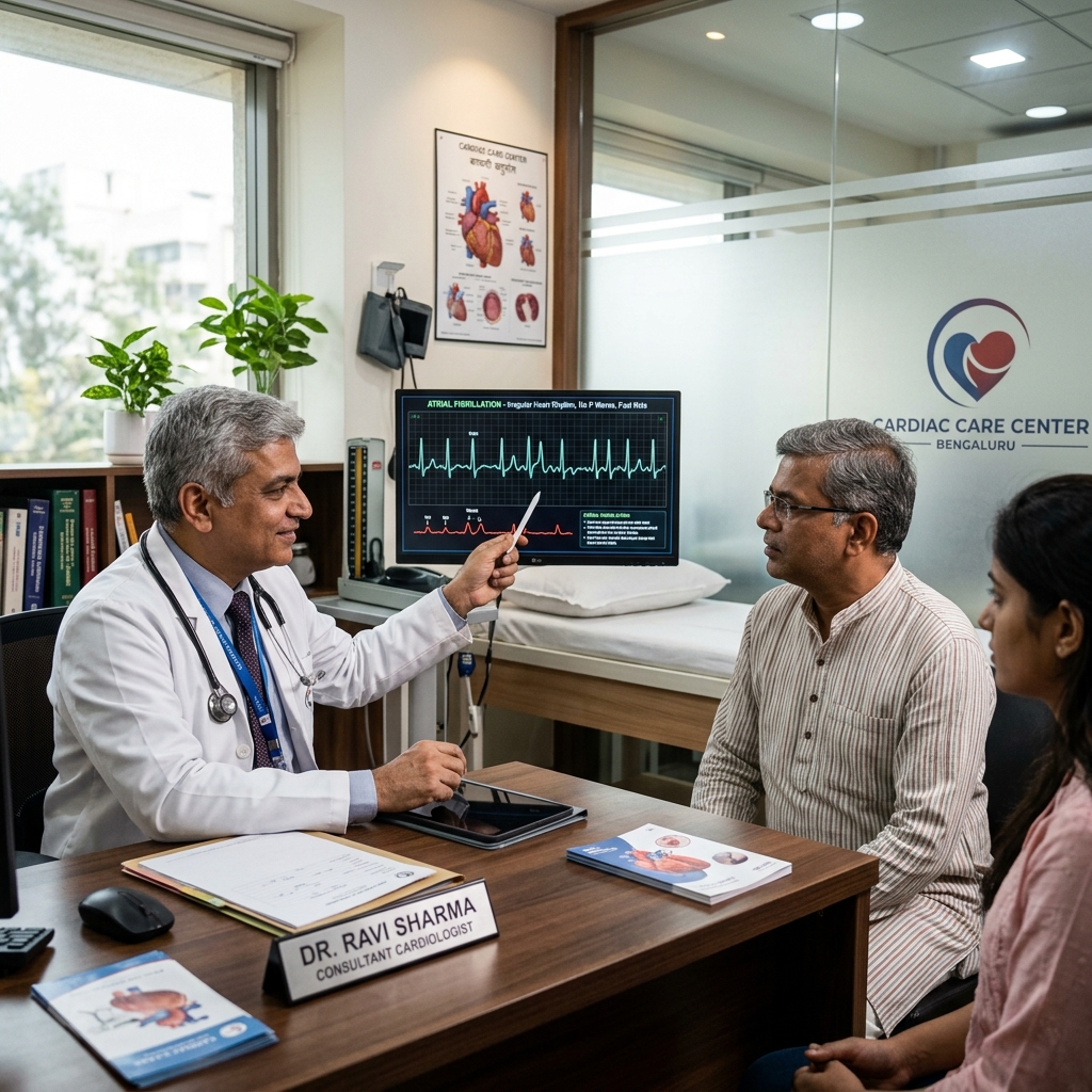
Atrial Fibrillation (AF)
ArrhythmiaAbsent P waves with irregularly irregular complexes. AF is the most common sustained arrhythmia and a leading preventable cause of stroke.
Left Bundle Branch Block (LBBB)
Conduction AbnormalityDelayed left ventricular conduction. New LBBB with symptoms requires rule-out of MI. Chronic LBBB with EF ≤35% warrants CRT device assessment.
Complete Heart Block (3rd Degree)
Urgent Action RequiredAtria and ventricles are electrically disconnected. Heart rate typically 20–40 bpm. High risk of cardiac arrest. Pacemaker required urgently.
Pathological Q Waves
Previous Heart AttackDeep Q waves indicate prior myocardial infarction (old heart attack). These are permanent markers of prior scar tissue in the heart muscle.
LV Hypertrophy (LVH)
Target Organ DamageIncreased QRS voltage indicating left ventricular hypertrophy. The heart's response to long-standing high blood pressure. Requires echocardiography for confirmation.
Preparation & Painless Procedure
Understand the minimal preparations required and the quick, comfortable process of a standard resting ECG.
No Fasting Required
Eat, drink, and take prescribed medications normally before your ECG. No special preparation is needed for this test.
Wear Appropriate Clothing
Wear a two-piece outfit that allows easy access to your chest, wrists, and ankles for electrode placement.
Avoid Lotions and Creams
Avoid body oils, greasy lotions, or heavy creams on your chest prior to electrode placement, as they interfere with signal quality.
Remove Metal Items
Remove metal items such as watches, necklaces, and bracelets if asked by the technician to avoid electrical interference.
The 5-Minute Trace
You will lie flat on a couch. Ten small electrodes are placed on your chest, arms, and legs. Real-time electrical readings are captured over 10 seconds. The trace is reviewed instantly and a printout is delivered immediately.
Risks of Ignoring ECG Abnormalities
Abnormal ECG findings that go uninvestigated can have serious consequences for patient outcomes.
Missed Heart Attack
ST elevation on ECG indicates an active heart attack. Every 30-minute delay in reperfusion increases mortality by approximately 7.5%. Immediate action is critical.
Undiagnosed Arrhythmia
Atrial fibrillation detected on ECG carries a 5-fold increased stroke risk if left untreated. Anticoagulation reduces this risk by approximately 65%.
Progressive Heart Block
First-degree heart block can progress to complete heart block without warning, causing syncope or cardiac arrest. Pacemaker implantation is life-saving.
Sudden Cardiac Death Risk
ECG findings such as long QT syndrome, Brugada pattern, or arrhythmogenic cardiomyopathy indicate elevated sudden death risk that requires specific intervention.
Treatment Based on ECG Findings
ECG results guide targeted treatment ranging from lifestyle modification to emergency revascularisation.
Emergency Reperfusion
STEMI on ECG triggers immediate transfer for primary angioplasty. The goal is door-to-balloon time under 90 minutes to salvage heart muscle.
Anticoagulation for AF
Atrial fibrillation detected on ECG requires risk stratification (CHA₂DS₂-VASc score) and anticoagulation to prevent stroke. Rate or rhythm control is then selected.
Pacemaker for Heart Block
Symptomatic bradycardia or high-degree AV block on ECG requires permanent pacemaker implantation. Dual-chamber pacing restores AV synchrony.
Ischaemia Management
ST depression or T wave inversion suggesting ischaemia triggers further testing (TMT, coronary angiography) and guideline-directed medical therapy including antiplatelets and statins.
Common Medications for ECG-Detected Conditions
ECG findings guide the selection of medications for rhythm control, ischaemia management, and risk reduction.
Lifestyle to Support Heart Health
Healthy lifestyle choices reduce the risk of developing conditions that produce abnormal ECG findings.
Heart-Healthy Diet
A Mediterranean-style diet rich in fruits, vegetables, whole grains, and healthy fats reduces cardiovascular risk and supports normal electrical function.
Regular Exercise
At least 150 minutes of moderate aerobic activity per week improves cardiovascular fitness and helps maintain normal heart rate and rhythm.
Avoid Smoking & Excess Alcohol
Tobacco use and excessive alcohol consumption are major risk factors for arrhythmias and ischaemic heart disease. Smoking cessation is the single most impactful lifestyle change.
Stress Management
Chronic stress and poor sleep can trigger palpitations and arrhythmias. Adequate sleep (7–8 hours) and stress reduction techniques are important for cardiac health.
ECG Guideline Standards
International guidelines define ECG interpretation standards and appropriate use criteria for screening.
When to See a Cardiologist
Certain ECG findings or symptoms warrant immediate or early cardiology consultation.
Chest Pain with ECG Changes
Chest pain or discomfort accompanied by ST changes on ECG requires emergency evaluation. Call emergency services or visit the nearest emergency department immediately.
Palpitations or Dizziness
Recurrent palpitations, lightheadedness, or fainting episodes warrant cardiology evaluation even with a normal resting ECG, as intermittent arrhythmias may require Holter monitoring.
Abnormal Screening ECG
If a routine ECG shows unexpected findings such as Q waves, bundle branch block, or LVH, a cardiology consultation is needed to determine the clinical significance.
Family History of Sudden Death
A family history of sudden cardiac death or inherited heart conditions requires cardiology screening including ECG, echocardiography, and possibly genetic testing.
Frequently Asked Questions
Detailed, clinical clarifications on resting electrocardiograms.
An ECG (electrocardiogram) is a 5-minute test that records the electrical activity of the heart from 12 different viewpoints using 10 electrodes on the chest, arms, and legs. It identifies the heart rate, rhythm, and any abnormalities in electrical conduction, including heart attacks (ST elevation), ischaemia (ST depression), arrhythmias (atrial fibrillation, heart block), conduction defects (bundle branch block), and chamber hypertrophy. No preparation is needed. The result is available immediately.
A 12-lead ECG can detect: heart rate and rhythm (normal sinus, AF, flutter, tachycardia, bradycardia); acute myocardial infarction (ST elevation — emergency); prior heart attack (pathological Q waves); ischaemia (ST depression, T wave inversion); bundle branch block (LBBB, RBBB); AV block (1st, 2nd, 3rd degree — complete heart block requires pacemaker); LV hypertrophy; QT prolongation; pericarditis; electrolyte disturbances; and drug toxicity effects.
Not necessarily. A normal ECG means the electrical activity of the heart appears normal at that moment — but significant coronary artery disease may produce no ECG changes at rest. Structural heart disease (valve abnormalities, cardiomyopathy) may not alter the ECG. Intermittent arrhythmias that are not present during the 10-second recording will not appear. A normal ECG is reassuring but does not exclude all cardiac conditions. Echocardiography, TMT, and Holter monitoring provide additional and complementary information.
An ECG records the heart's electrical activity — rhythm, conduction, and ischaemia. An echocardiogram (cardiac ultrasound) shows the physical structure of the heart — chamber sizes, wall thickness, pumping function (ejection fraction), and valve function. They provide completely different information. A patient can have a normal ECG but abnormal echocardiogram (valve disease, reduced EF), or a normal echo but abnormal ECG (arrhythmia, conduction block). Both tests are frequently ordered together.
A standard 12-lead ECG takes approximately 5 minutes from start to finish, with the actual recording lasting only 10–15 seconds. The test is completely painless. You may feel a slight cool sensation from the gel on the electrodes, and mild pressure when the stickers are applied to your chest. There are no needles, no radiation, and no recovery time. You can return to normal activities immediately after the test.
Yes, an ECG can show evidence of a prior heart attack through pathological Q waves — deep, wide deflections that represent scar tissue from a previous myocardial infarction. These Q waves are permanent markers that remain on the ECG for life. However, the ECG cannot tell you exactly when the heart attack occurred. A small heart attack or one affecting certain areas of the heart may not leave Q waves, so a normal ECG does not completely rule out a past cardiac event.
A stress ECG (Treadmill Stress Test or TMT) records your ECG continuously while you walk on a treadmill at progressively increasing speed and incline. This is different from a resting ECG because many coronary blockages only reduce blood flow during exertion when the heart demands more oxygen. A resting ECG can appear completely normal even with significant coronary artery disease. A stress ECG reveals ischaemic changes (ST depression) that appear only during exercise — providing information that a resting ECG cannot.
No special preparation is needed for a standard resting ECG. You can eat, drink, and take all your medications normally. We recommend wearing a two-piece outfit (shirt and pants or skirt) for easy chest access. Avoid applying body lotions or powders on your chest on the day of the test, as these can interfere with electrode adhesion. The entire procedure takes about 5 minutes and you can drive yourself home afterward.
“Advanced cardiovascular care. Restoring life, rhythm, and vitality.”

Dr. Amit Singh, FACC
Consultant Interventional Cardiologist
Related Tests & Conditions
ECG is often performed alongside other cardiac tests for a complete diagnostic evaluation.
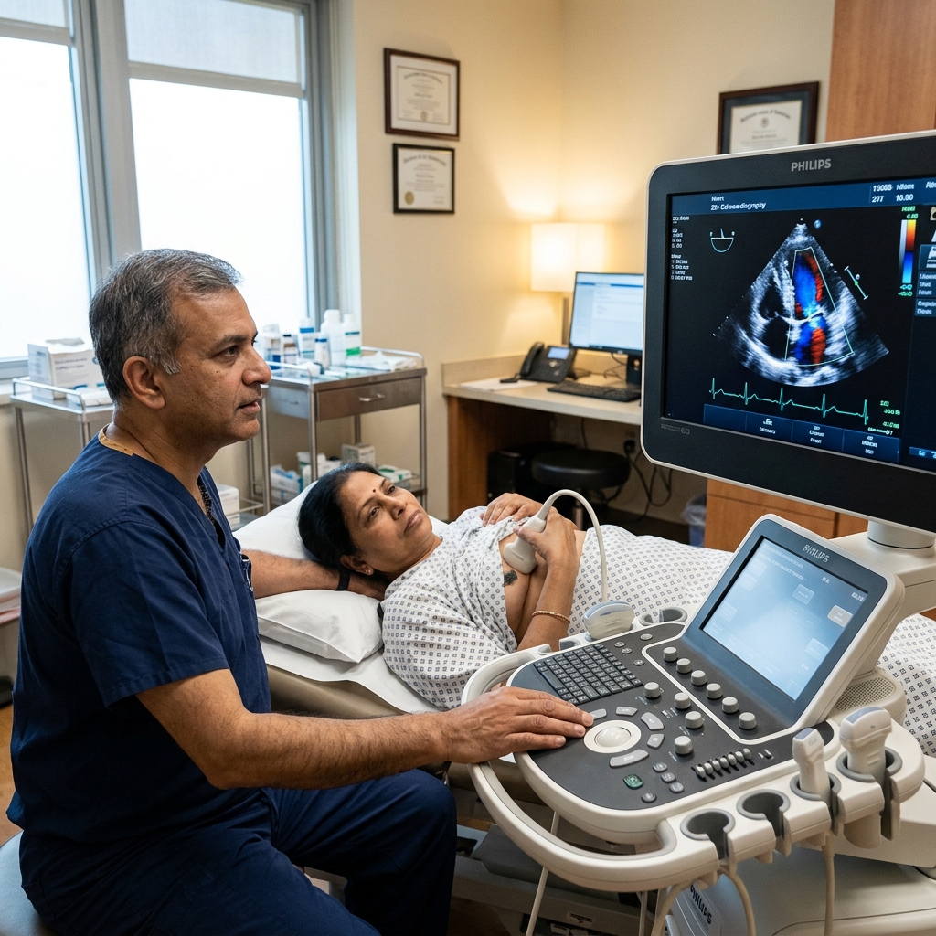
2D Echocardiography
Ultrasound imaging of heart structure, valves, and pumping function. Complements ECG by assessing structural abnormalities.
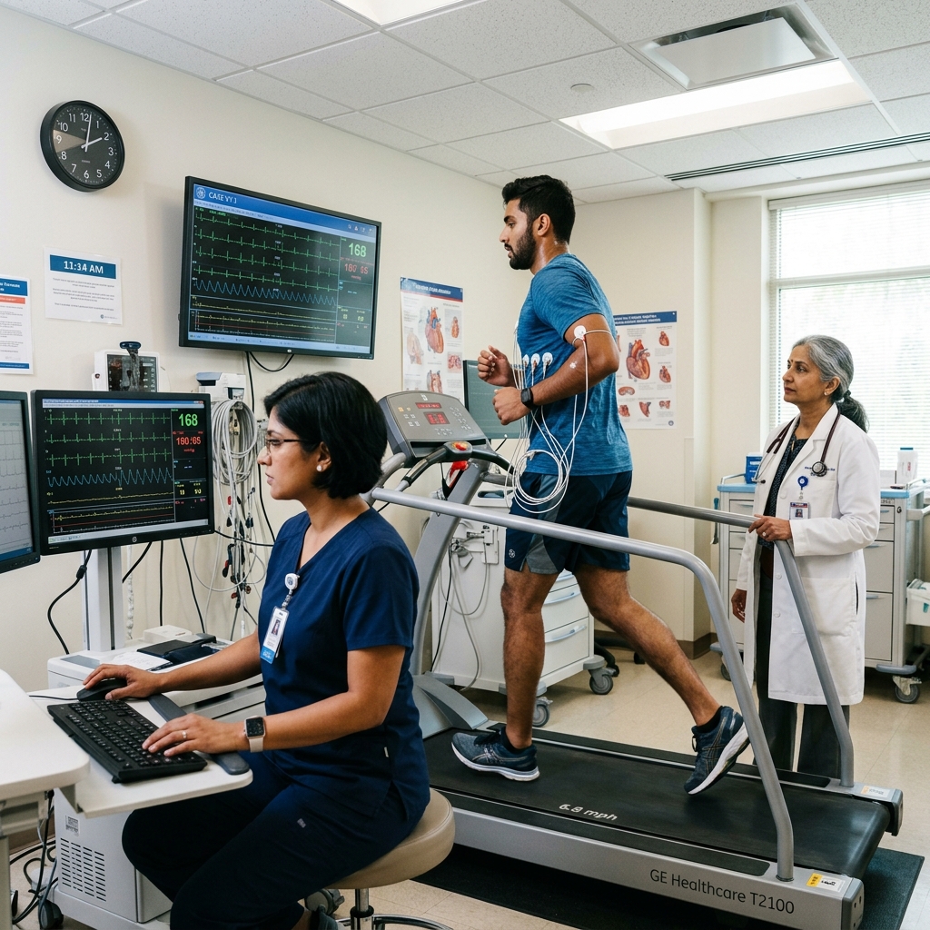
Treadmill Stress Test (TMT)
Exercise ECG to detect coronary artery disease not visible on resting ECG. Modified Bruce protocol with continuous monitoring.
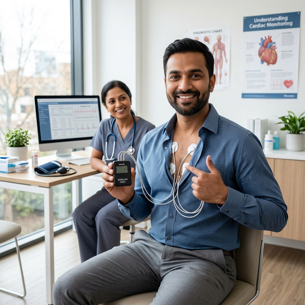
24-Hour Holter Monitoring
Continuous ambulatory ECG over 24–48 hours to capture intermittent arrhythmias that a resting 10-second ECG may miss.
Atrial Fibrillation
The most common sustained cardiac arrhythmia. ECG is essential for diagnosis and guiding stroke prevention therapy.
Book a Visit.
Pick a date and time that works for you.
Select a date
| Su | Mo | Tu | We | Th | Fr | Sa |
|---|---|---|---|---|---|---|
Book an Appointment with Dr. Amit Singh, FACC.
Schedule a cardiology consultation at Heartwise Clinic in Vashi, Navi Mumbai — online booking, WhatsApp, or call. Dr. Amit Singh offers in-clinic and secure video teleconsultations for patients across India and internationally.
Choose Date & Time
Pick a slot that fits your schedule from available morning or evening appointments.
Share Your Details
Provide your name, contact number, and a brief note about your cardiac concern or reason for visit.
Get Confirmed
Our clinical team confirms your slot within 24 hours via call or WhatsApp with pre-visit instructions.
Consult In-Clinic or Online
Visit Heartwise Clinic in Vashi or join a secure HD video teleconsultation from anywhere.
Consultation Options
In-Clinic Consultation
Kokilaben Hospital, Kopar Khairane & Heartwise Clinic, Vashi
HD Video Teleconsultation
Available pan-India and for international patients
WhatsApp Booking
Quick booking via +91 97695 17636 — reports & follow-ups
100+ appointments this month
Confirmed by our clinical team
4.9 / 5 rating
From patient reviews
Our Cardiology
Centers.
Dr. Amit Singh consults across multiple flagship centers and outreach clinics in Navi Mumbai & Dombivli to ensure specialized, top-tier cardiac care is directly accessible.
Navi Mumbai Sectors & Surrounding Nodes Served
Triple ESC & FACC Certified
International guidelines and clinical safety protocols applied across all heart centers.
“Beat Better. Live Wiser.”
Dr. Amit Singh, FACC
Consultant Interventional Cardiologist
Medical Disclaimer: This article has been written and reviewed by Dr. Amit Singh, FACC, for educational purposes only. It does not constitute personalised medical advice and should not be used as a substitute for a consultation with a qualified cardiologist. Individual clinical decisions must be made by a treating physician based on complete medical history and examination. If you are experiencing chest pain, breathlessness, or other cardiac symptoms, seek emergency medical care immediately.