
Rhythm & Palpitations
Palpitations — the awareness of your heartbeat as racing, thumping, or irregular — are one of the most common cardiac presentations. Systematic Holter tracking is crucial.
What Is Causing Your Palpitations?
Benign extra beats are present in virtually all humans, but rapid or irregular fluttering must be stratified.
Palpitations range from harmless premature beats (ectopics) to complex tachyarrhythmias (Atrial Fibrillation, SVT, or VT) and slow blockages (Sick Sinus Syndrome). Early classification is key.
Causes & Risk Factors for Palpitations
Palpitations arise from cardiac electrical abnormalities, systemic conditions, or lifestyle triggers.
Ectopic Beats (PVCs / PACs)
Premature contractions produce a 'missed beat' or 'flip-flop' sensation. Present in virtually all people. Isolated low-burden PVCs in a structurally normal heart require no treatment.
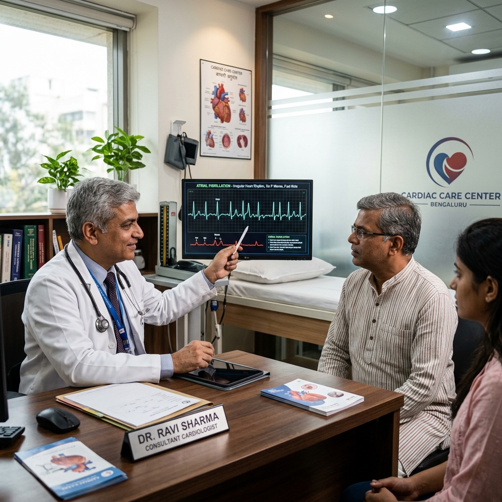
Atrial Fibrillation
Irregularly irregular palpitations — may feel like 'chaotic fluttering.' 5× increased stroke risk. Requires CHA₂DS₂-VASc scoring and anticoagulation.
Supraventricular Tachycardia (SVT)
Sudden onset racing heart at 150–250 bpm — typically paroxysmal, starts and stops abruptly. Distressing but rarely dangerous in structurally normal hearts.
Ventricular Tachycardia (VT)
Fast regular rhythm from the ventricle — 120–200 bpm. Associated with structural heart disease. Risk of degeneration to VF and sudden cardiac death.
Sick Sinus Syndrome / Heart Block
Sinus node dysfunction causes slow rates, pauses, or alternating brady-tachy. Complete heart block is life-threatening — ventricle escapes at 20–40 bpm.
Non-Cardiac Causes
Anxiety, panic attacks, anaemia, thyrotoxicosis, fever, and caffeine excess can all cause awareness of a fast but normal sinus rhythm. The heart is structurally and electrically normal.
Classification of Common Heart Rhythm Disorders
We stratify palpitations by clinical risk and present guideline-recommended therapies.
Signs & Symptoms of Arrhythmia
Symptoms vary by arrhythmia type, duration, and underlying heart structure.
Palpitations
Sensation of racing, thumping, fluttering, or irregular heartbeat. May be felt in the chest, throat, or neck. Can be episodic or sustained.
Dizziness & Presyncope
Lightheadedness or feeling faint, especially during rapid arrhythmias or prolonged pauses. Reduced cerebral perfusion during tachycardia or bradycardia.
Shortness of Breath
Reduced cardiac output during arrhythmia causes breathlessness. May be exertional or occur at rest during sustained episodes.
Syncope (Fainting)
Sudden loss of consciousness from profound bradycardia, tachycardia (VT), or asystole. Requires urgent investigation.
Chest Discomfort
Chest pain or pressure during rapid heart rates may indicate ischaemia from supply-demand mismatch, especially with underlying CAD.
Fatigue & Reduced Capacity
Chronic arrhythmias like persistent AF cause fatigue from reduced cardiac output. Patients often attribute it to aging.
Our Diagnostic Pathway for Heart Rhythm Issues
Standard cardiac screening to record electrical events across 100,000+ continuous heartbeats.
Resting 12-Lead ECG
First test. Captures AF (irregularly irregular baseline, absent P waves), SVT (narrow regular tachycardia), VT (wide complex tachycardia), or heart block.
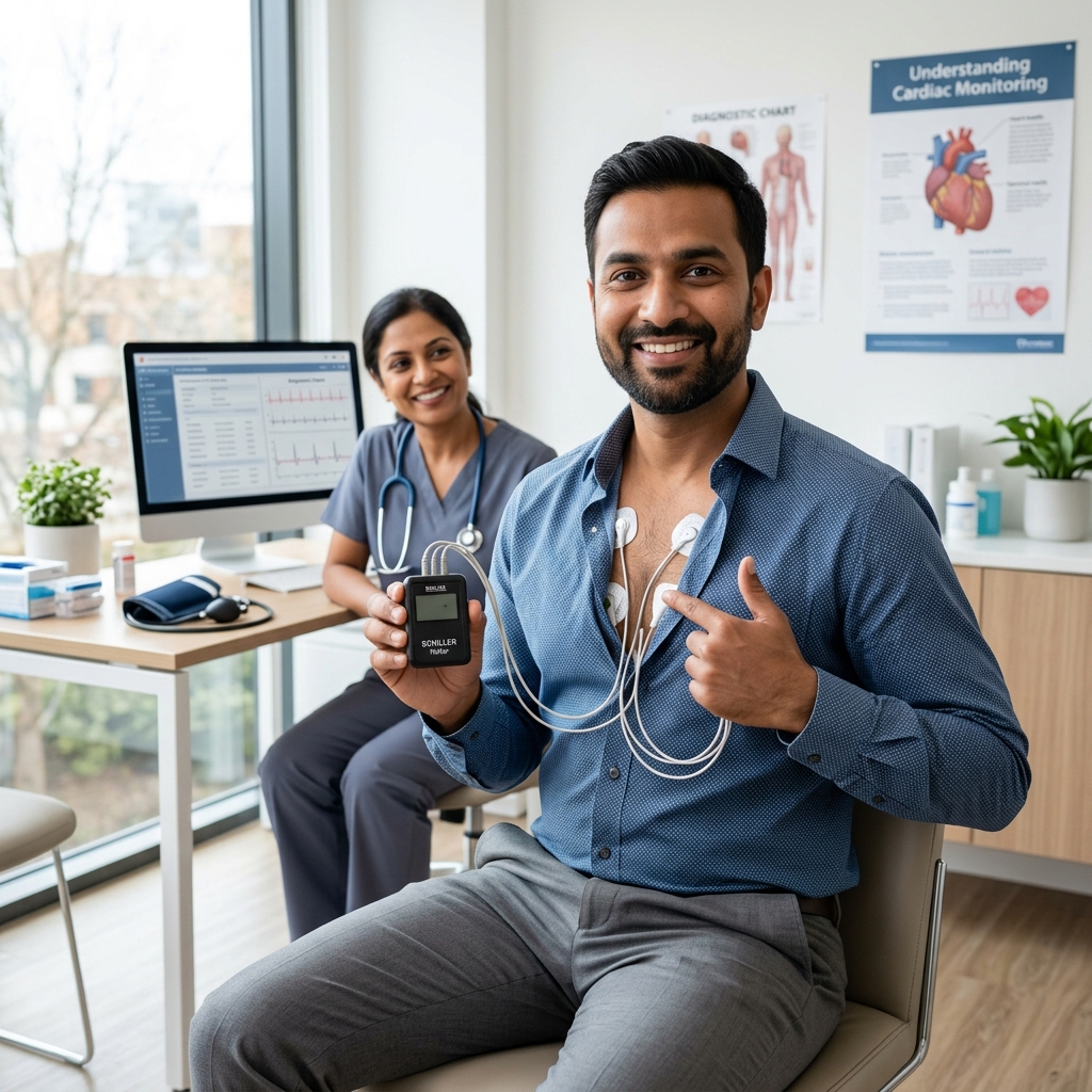
24-Hour Holter Monitoring
Continuous ECG for 24–48 hours captures arrhythmias between episodes. Symptom diary correlation confirms whether palpitations coincide with ECG changes.
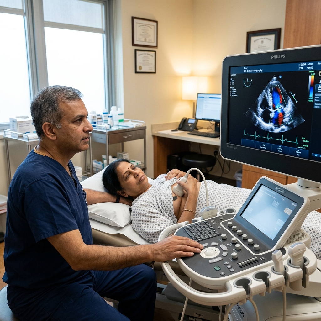
2D Echocardiography
Assesses ejection fraction (critical — VT with reduced EF carries ICD indication), left atrial dilation (AF substrate), HOCM, and valve disease.
Extended Monitoring (7-day / ILR)
If standard tests are normal but episodes are infrequent, 7-day patch monitoring or implantable loop recorder (ILR) detects paroxysmal AF.
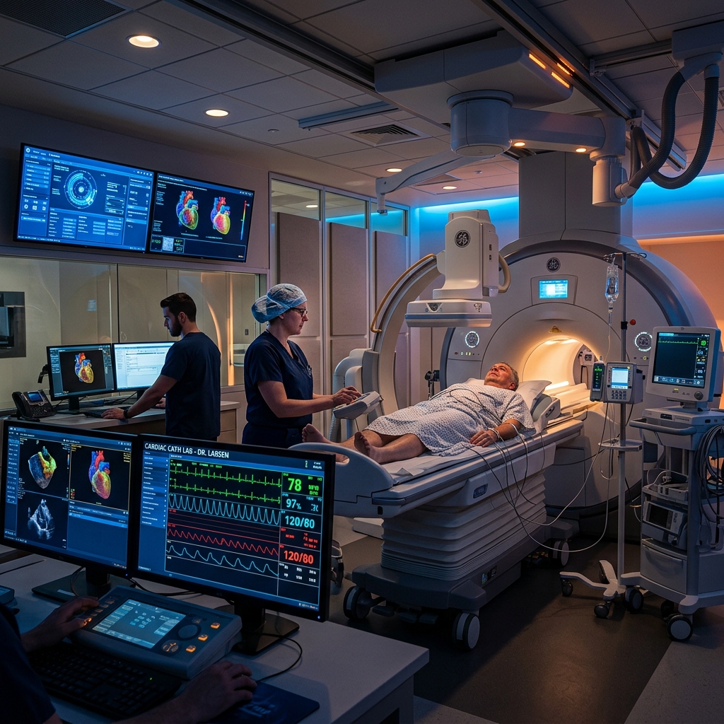
Electrophysiology Study (EPS)
Invasive catheter-based mapping of the heart's electrical system. Definitive diagnosis for SVT, VT, and syncope of unknown origin. Guides ablation therapy.
What Happens If Left Untreated?
Depending on the type, untreated arrhythmias can lead to stroke, heart failure, or sudden cardiac death.
Stroke (from AF)
Untreated AF with stroke risk factors leads to a 5× increased stroke risk. Cardioembolic strokes from AF are large, disabling, and often fatal.
Tachycardia-Mediated Cardiomyopathy
Chronic uncontrolled tachycardia (AF, atrial tachycardia, VT) can cause reversible LV dysfunction and heart failure.
Sudden Cardiac Death (from VT/VF)
Sustained VT can degenerate into ventricular fibrillation — a lethal arrhythmia requiring immediate defibrillation. ICD prevents this in high-risk patients.
Syncope & Falls
Complete heart block or prolonged pauses cause syncope. Falls from syncope can cause serious injury, particularly in the elderly.
Treatment Options for Palpitations & Arrhythmia
Treatment is tailored to the specific arrhythmia, symptom burden, and underlying heart disease.
Lifestyle Modification & Reassurance
For benign ectopics: reduce caffeine and alcohol, optimise sleep, manage stress. Reassurance that isolated PVCs in a normal heart are harmless.
Antiarrhythmic Medications
Beta-blockers (bisoprolol) for symptomatic PVCs and rate control. Flecainide or propafenone for SVT. Amiodarone for VT. Dronedarone for AF maintenance.
Catheter Ablation
Curative procedure for SVT (95% success). Pulmonary vein isolation for AF. Substrate ablation for VT. Performed via venous access under conscious sedation.
Pacemaker & ICD Implantation
Pacemaker for symptomatic bradycardia or heart block. ICD for VT/VF prevention in high-risk patients (EF ≤35%, prior VT arrest).
Medications for Arrhythmia
Antiarrhythmic drugs are classified by their mechanism of action and clinical indication.
Lifestyle Changes & Self-Care for Palpitations
Simple measures can reduce the frequency and severity of palpitation episodes.
Avoid Triggers
Reduces episode frequencyCaffeine, alcohol, nicotine, and recreational drugs are common triggers. Keep a symptom diary to identify personal triggers.
Optimise Sleep
Improves autonomic balanceSleep deprivation and shift work disturb cardiac autonomic balance and increase arrhythmia susceptibility. Aim for 7–8 hours per night.
Vagal Manoeuvres
First-line for SVT acutelyFor sudden regular racing (SVT), vagal stimulation like bearing down or applying cold water to the face can terminate the circuit instantly.
Stress Management
Reduces symptom perceptionAnxiety and panic attacks amplify palpitation perception. Relaxation techniques, deep breathing, and CBT reduce symptom burden.
Track Your Pulse
Aids diagnosisCheck your wrist pulse immediately during episodes. Note rate, regularity, and duration. Timestamp logs help correlate with Holter findings.
Stay Hydrated
Simple preventive measureDehydration can trigger palpitations. Maintain adequate fluid intake, especially in hot weather and during exercise.
Guidelines & Latest Evidence
Current guidelines inform the management of cardiac arrhythmias.
When to See a Doctor
Certain palpitation characteristics require prompt medical evaluation.
Palpitations with Syncope or Near-Syncope
Fainting or severe lightheadedness during palpitations suggests a dangerous arrhythmia requiring immediate evaluation.
Rapid Heart Rate >150 bpm
A sustained heart rate above 150 bpm at rest, especially of sudden onset, requires assessment to diagnose and treat the arrhythmia.
Palpitations with Chest Pain or Breathlessness
Combined palpitations with chest pain or shortness of breath may indicate ischaemia, HF, or life-threatening arrhythmia.
Known Structural Heart Disease with New Palpitations
Patients with prior MI, heart failure, cardiomyopathy, or valve disease should not ignore new palpitations — VT risk is higher.
Frequently Asked Questions
Guideline-directed clarifications regarding extra beats and pacing requirements.
Palpitations are caused by: benign ectopic beats (premature atrial or ventricular contractions — the most common cause); atrial fibrillation (irregularly irregular rhythm — most important to exclude due to stroke risk); supraventricular tachycardia or SVT (sudden racing heart at 150–250 bpm, typically paroxysmal); ventricular tachycardia (dangerous — associated with structural heart disease); sinus tachycardia from anxiety, anaemia, thyrotoxicosis, or fever; and sick sinus syndrome or heart block (slow rate causing bradycardia sensation). ECG and Holter monitoring are required to identify the cause.
Most palpitations are benign — caused by ectopic beats, anxiety, or caffeine in a structurally normal heart. However, some arrhythmias carry real risk: atrial fibrillation (5× increased stroke risk — requires anticoagulation); ventricular tachycardia (risk of sudden cardiac death, especially with structural heart disease); and complete heart block (may cause syncope and require pacemaker). The distinction between benign and dangerous requires ECG, Holter monitoring, and echocardiography — not clinical assessment alone.
See a cardiologist promptly if palpitations are accompanied by breathlessness, chest pain, or dizziness; cause fainting (syncope) or near-fainting; feel very rapid (>150 bpm); are irregular rather than just fast; occur during exercise; occur after age 40; or occur in a patient with known structural heart disease (heart failure, prior MI, cardiomyopathy, or family history of sudden cardiac death). Palpitations in a healthy young person without structural disease are more likely benign, but still require ECG and Holter before reassurance can be given.
The standard diagnostic workup for palpitations begins with a resting 12-lead ECG to capture any arrhythmia at the time of evaluation. If the resting ECG is normal, a 24-hour Holter monitor is the next step — it records every heartbeat over a full day and night, allowing correlation of symptoms with actual rhythm. If palpitations are infrequent, extended monitoring with a 7-day patch recorder or an implantable loop recorder (ILR) may be used. A 2D echocardiogram is always performed to assess structural heart disease, ejection fraction, and valve function. Blood tests to check thyroid function, anaemia, and electrolyte imbalances complete the evaluation.
Yes — both caffeine and alcohol are well-known triggers for palpitations. Caffeine is a stimulant that increases heart rate and can provoke ectopic beats (PVCs and PACs) in susceptible individuals. The effect varies widely — some people experience palpitations after a single cup of coffee while others tolerate multiple cups. Alcohol, particularly binge drinking, can precipitate atrial fibrillation — known as 'holiday heart syndrome' — as well as other arrhythmias. Both substances can also worsen existing arrhythmias such as AF or SVT. Reducing or eliminating caffeine and alcohol is a simple first step in managing palpitations. If symptoms persist despite avoidance, formal cardiac evaluation is needed.
Palpitations are common during pregnancy due to the significant physiological changes — increased blood volume (up to 50%), elevated heart rate (10–20 bpm above baseline), hormonal shifts, and the mechanical effects of the growing uterus on venous return. Most palpitations in pregnancy are benign and caused by increased awareness of normal sinus tachycardia or benign ectopic beats. However, pregnancy can also unmask or worsen underlying arrhythmias such as SVT or AF. Any palpitations associated with chest pain, breathlessness, dizziness, or fainting require urgent cardiac evaluation. ECG and Holter monitoring are safe during pregnancy. Most antiarrhythmic medications can be used with appropriate precautions after cardiology and obstetric consultation.
Palpitations are the sensation of feeling your heartbeat — whether it feels fast, pounding, fluttering, or irregular. They are a symptom, not a diagnosis. An arrhythmia is an actual abnormality of the heart's electrical rhythm — such as atrial fibrillation, supraventricular tachycardia, or ventricular tachycardia — that can be documented on ECG or Holter monitoring. Not all palpitations are caused by arrhythmias — increased awareness of a normal heartbeat (sinus tachycardia) due to anxiety, anaemia, or fever also causes palpitations without any rhythm disorder. The distinction between a benign symptom and a clinically significant arrhythmia requires ECG documentation, which is why 24-hour Holter monitoring is the standard diagnostic test.
Yes — stress and anxiety are among the most common causes of palpitations. The sympathetic nervous system response to stress releases adrenaline, which increases heart rate and the force of contraction, and can trigger ectopic beats (premature ventricular or atrial contractions). Panic attacks can produce heart rates of 120–160 bpm, along with chest tightness, hyperventilation, and a sense of impending doom — closely mimicking dangerous arrhythmias. However, it is critical to complete a cardiac evaluation before attributing palpitations to anxiety, as the symptoms of VT, SVT, and AF can be identical. Once cardiac causes are excluded, stress management, cognitive behavioural therapy, and relaxation techniques can significantly reduce symptom burden.
“Advanced cardiovascular care. Restoring life, rhythm, and vitality.”

Dr. Amit Singh, FACC
Consultant Interventional Cardiologist
Cardiac Services Available at Heartwise
Dr. Amit Singh offers comprehensive cardiac diagnostics and management for this condition at Heartwise Cardiology Clinic, Vashi.
24-Hour Holter
Continuous ECG monitoring capturing ectopics, SVT, and paroxysmal AF.
Resting ECG
Immediate 12-lead ECG to identify baseline arrhythmias and conduction defects.
2D Echocardiography
Structural heart assessment — EF, LA size, and valve disease evaluation.
Atrial Fibrillation
Comprehensive AF management including rhythm control and anticoagulation.
Book a Visit.
Pick a date and time that works for you.
Select a date
| Su | Mo | Tu | We | Th | Fr | Sa |
|---|---|---|---|---|---|---|
Book an Appointment with Dr. Amit Singh, FACC.
Schedule a cardiology consultation at Heartwise Clinic in Vashi, Navi Mumbai — online booking, WhatsApp, or call. Dr. Amit Singh offers in-clinic and secure video teleconsultations for patients across India and internationally.
Choose Date & Time
Pick a slot that fits your schedule from available morning or evening appointments.
Share Your Details
Provide your name, contact number, and a brief note about your cardiac concern or reason for visit.
Get Confirmed
Our clinical team confirms your slot within 24 hours via call or WhatsApp with pre-visit instructions.
Consult In-Clinic or Online
Visit Heartwise Clinic in Vashi or join a secure HD video teleconsultation from anywhere.
Consultation Options
In-Clinic Consultation
Kokilaben Hospital, Kopar Khairane & Heartwise Clinic, Vashi
HD Video Teleconsultation
Available pan-India and for international patients
WhatsApp Booking
Quick booking via +91 97695 17636 — reports & follow-ups
100+ appointments this month
Confirmed by our clinical team
4.9 / 5 rating
From patient reviews
Our Cardiology
Centers.
Dr. Amit Singh consults across multiple flagship centers and outreach clinics in Navi Mumbai & Dombivli to ensure specialized, top-tier cardiac care is directly accessible.
Navi Mumbai Sectors & Surrounding Nodes Served
Triple ESC & FACC Certified
International guidelines and clinical safety protocols applied across all heart centers.
“Beat Better. Live Wiser.”
Dr. Amit Singh, FACC
Consultant Interventional Cardiologist
Medical Disclaimer: This article has been written and reviewed by Dr. Amit Singh, FACC, for educational purposes only. It does not constitute personalised medical advice and should not be used as a substitute for a consultation with a qualified cardiologist. Individual clinical decisions must be made by a treating physician based on complete medical history and examination. If you are experiencing chest pain, breathlessness, or other cardiac symptoms, seek emergency medical care immediately.