ASD Device Closure Hole in the Heart — Closed Without Open Surgery
An Atrial Septal Defect (ASD) — commonly called a “hole in the heart” — is closed via collapsible metallic mesh deployed through a femoral catheter. Enjoy a 95%+ success rate, TOE-guided safety, and a rapid 24–48 hour recovery.
Understanding Atrial Septal Defects (ASD)
An evidence-based overview of interatrial shunts, right ventricular volume overload, and clinical indications.
An atrial septal defect (ASD) is an abnormal opening in the interatrial septum — the wall separating the right and left atria. Because left atrial pressure is higher, blood flows left-to-right through the defect, overloading the right heart and pulmonary circulation. The shunt severity is quantified by the Qp:Qs ratio (pulmonary to systemic blood flow). Significant shunting (Qp:Qs ≥ 1.5:1) causes right heart enlargement, right heart failure over time, and pulmonary arterial hypertension if untreated for decades. Most patients are asymptomatic in childhood and young adulthood. Symptoms typically emerge in the third or fourth decade: breathlessness, reduced exercise tolerance, palpitations, and occasionally paradoxical embolism causing stroke (a clot from the right heart crosses through the ASD into the arterial circulation). This is why cryptogenic stroke (stroke with no identified cause) triggers investigation for PFO or ASD — and closure to prevent recurrence. The CLOSE trial (NEJM 2017) demonstrated PFO closure reduces stroke recurrence versus medical therapy alone in patients under 60 with cryptogenic stroke.
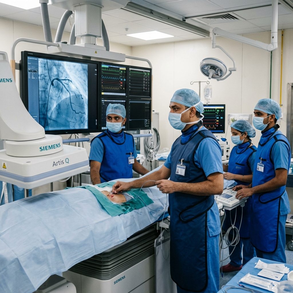
The Device Closure Procedural Steps
A comprehensive clinical breakdown of how the nitinol mesh septal occluder is positioned and expanded.
Echocardiographic Sizing & Sedation
Conducted in the Cath Lab under general anesthesia or deep conscious sedation. A transoesophageal echocardiography (TOE) probe is introduced to take precise 2D/3D measurements of the defect diameter and evaluate the surrounding rims to select the ideal device diameter.
Femoral Sheath Access
A small catheter sheath is placed in the femoral vein in the groin. No arterial access or large incisions are needed. Heparin is administered intravenously to prevent clot formation during the procedure.
Crossing the Interatrial Hole
Under real-time fluoroscopy (X-ray) and TOE guidance, a soft guidewire and catheter are advanced through the femoral vein, up into the right atrium, and directly across the septal hole into the left atrium.
Mesh Occluder Deployment
A self-expanding, double-disc nitinol mesh device (Amplatzer or equivalent) is compressed inside a delivery catheter. The left atrial disc is deployed first, pulled snug against the septum, followed by the central waist and right atrial disc, sandwiching the defect.
Stability Verification (Minnesota Wiggle)
Before final release, the cardiologist performs a gentle push-pull test (Minnesota Wiggle) under fluoroscopy. TOE confirms the device is securely anchored, creates a complete seal, and does not impinge on surrounding valves or pulmonary veins.
Release & Hemostasis
The device is unscrewed from the delivery cable. Sheaths are removed, and a pressure bandage is applied to the groin. The patient is monitored in the recovery suite overnight and routinely discharged the following morning.
Procedural Benefits & Risks
Reviewing the evidence-based profile of transcatheter closure compared to surgical equivalents.
Core Clinical Benefits
- Avoidance of sternotomy or cardiopulmonary bypass circuits
- Highly predictable >95% procedural success in suitable anatomy
- Groin access hemostasis allows rapid next-day discharge
- Fast return to physical activities within 1–2 weeks
- Relieves right heart volume overload, leading to reverse ventricular remodeling
- Significantly reduces risk of paradoxical stroke or systemic emboli
Potential Risks & Incidence
- Device Embolisation: <0.5% — Catheter-retrievable in most cases
- New Atrial Arrhythmia: 3–5% — Usually self-limiting in the first 3 months
- Residual Shunting: 5–10% — Small micro-leaks typically seal over 6 months
- Late Device Erosion: <0.1% — Extremely rare complication with proper sizing
- Groin Access Hematoma: <1% — Managed conservatively with compression
Frequently Asked Questions
Detailed, peer-reviewed answers to the most common patient concerns regarding stenting and long-term care.
An atrial septal defect (ASD) is a hole in the wall (septum) between the right and left upper chambers of the heart (atria). Blood shunts left-to-right through the hole, overloading the right heart. Most patients are asymptomatic for decades before developing breathlessness, palpitations, or right heart failure. ASD is one of the most common adult congenital heart conditions. Transcatheter device closure is the preferred treatment for suitable secundum-type ASDs.
Transcatheter ASD closure uses a collapsible metallic mesh device — delivered via a catheter through the femoral vein — to permanently close the hole between the atria, without opening the chest. The device is guided by transoesophageal echocardiography (TOE). Closure success exceeds 95% for suitable secundum-type defects. Hospital stay is 24–48 hours. The procedure is preferred over surgical closure per ACC/AHA and ESC guidelines (Class I recommendation).
Untreated significant ASD causes progressive right heart enlargement, right heart failure, atrial arrhythmias, pulmonary arterial hypertension, and paradoxical embolism causing stroke. In a small proportion, Eisenmenger syndrome — irreversible pulmonary hypertension with reversed shunting — can develop, making closure impossible. Closure before right heart dilation or pulmonary hypertension produces the best outcomes.
Very small ASDs in infants (less than 5–6 mm) may close spontaneously in the first 2 years of life. After age 2, spontaneous closure is rare. Moderate to large ASDs do not close on their own. Adults presenting with ASD have persistent defects requiring intervention. Transcatheter device closure is preferred for suitable secundum-type defects.
ASD device closure has a major complication rate below 1–2% at experienced centres. Rare risks include device embolisation (<0.5%, retrievable), new atrial arrhythmia in first weeks (3–5%, usually transient), small residual shunt (5–10%, most seal within 6 months), and the very rare late complication of device erosion (<0.1%, related to oversizing). Patients require 6 months of antiplatelet therapy while the device endothelialises.
Who Needs ASD Closure?
Indications for transcatheter ASD closure based on shunt severity, anatomy, and clinical presentation.
Transcatheter ASD Closure Device Types
Overview of the self-expanding double-disc nitinol mesh occluder used for transcatheter ASD closure.
Transcatheter ASD Closure Device
Nitinol mesh with PET fabric — self-expanding double-disc design
Technical Specs
- •Size range: 4–40 mm diameter
- •Delivery sheath: 8–14 French
- •Nitinol frame with PET fabric inserts
- •Retrievable and repositionable before release
- •MRI-conditional after endothelialisation (3 months)
Device Closure vs Surgical Closure
Head-to-head comparison of transcatheter device closure versus open surgical ASD repair.
Pre-ASD Closure Workup & Preparation
Essential investigations and patient preparation before transcatheter ASD device closure.
ASD Closure Evidence Base
Key studies supporting transcatheter device closure outcomes, safety, and long-term efficacy.
Transcatheter device closure has >95% success rate for secundum ASDs with major complication rate <2%
O'Byrne ML et al. JACC 2019;73:1530–42Closure eliminates right-to-left shunt, prevents paradoxical embolism, and reduces long-term PAH risk
ESC Guidelines — CHD 2020 updateFreedom from device-related adverse events at 5 years in 98% of patients with Amplatzer-type devices
Haas NA et al. EuroIntervention 2020;16Post-ASD Closure Results Interpretation
Key echocardiographic and haemodynamic parameters for assessing ASD closure success.
Post-Closure Care & Long-Term Support
Ensuring device safety and endothelialization over the critical six-month recovery timeline.
In Hospital (24–48 Hours)
- Bed rest 4–6 hours after sheath removal; gradual mobilisation
- Chest X-ray and echo within 24 hours to confirm device position and exclude pericardial effusion
- Discharge typically next day after confirming stable device position and no complications
First Month at Home
- Antiplatelet therapy: aspirin + clopidogrel for 3–6 months while device endothelialises
- No heavy lifting for 2 weeks; avoid contact sports for 4 weeks
- Wound care: keep femoral access site dry for 48 hours
- Endocarditis prophylaxis for dental procedures in first 6 months
- Report any palpitations, chest pain, breathlessness, or fever immediately
Long-Term Follow-Up
- Echocardiogram at 1 month, 6 months, 12 months, then every 3–5 years
- No activity restrictions after 6 months once endothelialisation complete
- Lifelong echo surveillance for device erosion risk (rare, <0.1%)
- MRI safe after 3 months with MRI-conditional devices
Favourable vs Unfavourable Profiles for ASD Closure
Detailed breakdown of patient characteristics that determine suitability for transcatheter device closure.
Favourable Profile
- Secundum-type ASD with adequate rims (≥5mm)
- Moderate to large ASD with significant shunt (Qp:Qs ≥1.5:1)
- RV dilation or volume overload on echo
- Paradoxical embolism / cryptogenic stroke with PFO/ASD
- Symptomatic (dyspnoea, fatigue, palpitations, exercise intolerance)
Less Favourable
- Primum-type or sinus venosus ASD (requires surgery)
- Secundum ASD with deficient rims (<5mm) — unsuitable for device stability
- ASD >38mm or multiple fenestrations
- Eisenmenger syndrome with fixed PAH and right-to-left shunt
- Active endocarditis or untreated sepsis
- Intracardiac thrombus or recent pulmonary embolism
“Precision in structural interventions. Excellence in clinical outcomes.”

Dr. Amit Singh, FACC
Consultant Interventional Cardiologist
Cardiac Services Available at Heartwise
Dr. Amit Singh offers comprehensive cardiac diagnostics and management at Heartwise Cardiology Clinic, Vashi.
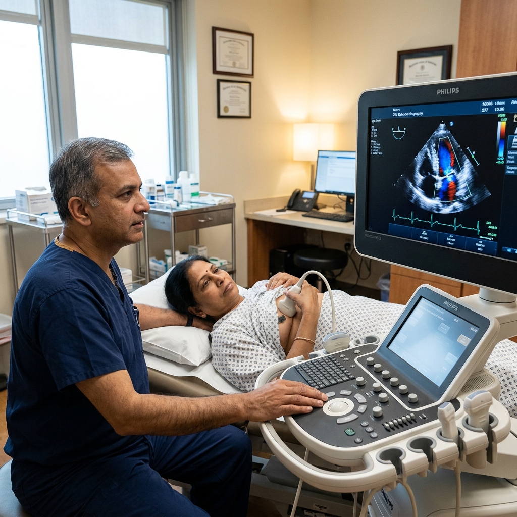
2D Echocardiography
Detailed echo assessment including TOE for ASD sizing and planning.

Resting 12-Lead ECG
Baseline ECG to assess rhythm and conduction.

Cardiac Risk Assessment
Pre-procedure risk stratification and cardiac evaluation.
Heart Failure
Long-term follow-up for LV function after ASD closure.
Book a Visit.
Pick a date and time that works for you.
Select a date
| Su | Mo | Tu | We | Th | Fr | Sa |
|---|---|---|---|---|---|---|
Book an Appointment with Dr. Amit Singh, FACC.
Schedule a cardiology consultation at Heartwise Clinic in Vashi, Navi Mumbai — online booking, WhatsApp, or call. Dr. Amit Singh offers in-clinic and secure video teleconsultations for patients across India and internationally.
Choose Date & Time
Pick a slot that fits your schedule from available morning or evening appointments.
Share Your Details
Provide your name, contact number, and a brief note about your cardiac concern or reason for visit.
Get Confirmed
Our clinical team confirms your slot within 24 hours via call or WhatsApp with pre-visit instructions.
Consult In-Clinic or Online
Visit Heartwise Clinic in Vashi or join a secure HD video teleconsultation from anywhere.
Consultation Options
In-Clinic Consultation
Kokilaben Hospital, Kopar Khairane & Heartwise Clinic, Vashi
HD Video Teleconsultation
Available pan-India and for international patients
WhatsApp Booking
Quick booking via +91 97695 17636 — reports & follow-ups
100+ appointments this month
Confirmed by our clinical team
4.9 / 5 rating
From patient reviews
Our Cardiology
Centers.
Dr. Amit Singh consults across multiple flagship centers and outreach clinics in Navi Mumbai & Dombivli to ensure specialized, top-tier cardiac care is directly accessible.
Navi Mumbai Sectors & Surrounding Nodes Served
Triple ESC & FACC Certified
International guidelines and clinical safety protocols applied across all heart centers.
“Beat Better. Live Wiser.”
Dr. Amit Singh, FACC
Consultant Interventional Cardiologist
Medical Disclaimer: This article has been written and reviewed by Dr. Amit Singh, FACC, for educational purposes only. It does not constitute personalised medical advice and should not be used as a substitute for a consultation with a qualified cardiologist. Individual clinical decisions must be made by a treating physician based on complete medical history and examination. If you are experiencing chest pain, breathlessness, or other cardiac symptoms, seek emergency medical care immediately.