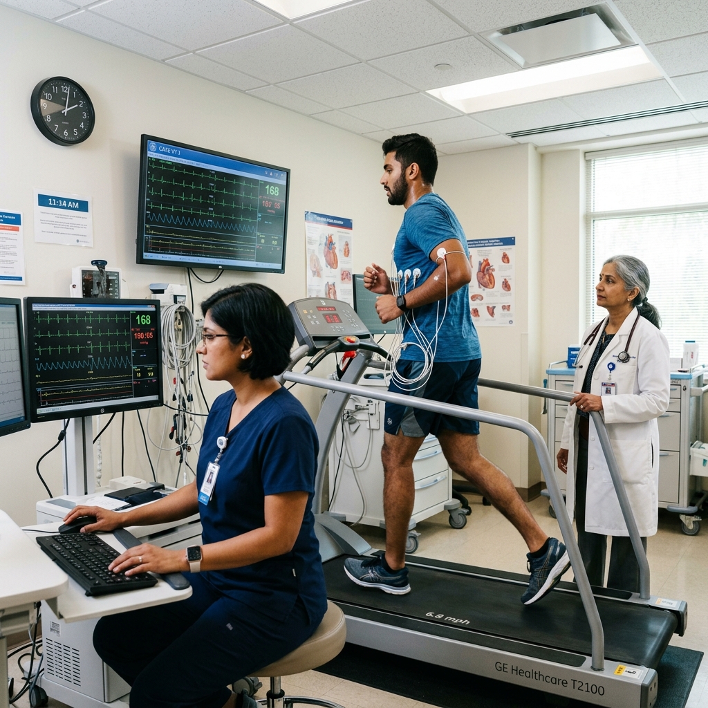
Treadmill Stress Test (TMT)
A Treadmill Stress Test (TMT) records a continuous 12-lead ECG while walking under a graded protocol. This reveals potential blood flow restriction (ischaemia) to the heart muscles during physical exertion, which often remains hidden at rest.
The Science of Cardiac Stress Testing
When at rest, narrowing of the coronary arteries may produce no symptoms and a normal ECG. Graded exercise reveals latent arterial blockages.
By progressively increasing speed and treadmill grade, the test drives up cardiac work, forcing the heart to draw more blood. A significant blockage (>70%) restricts the flow of oxygenated blood. This relative oxygen deficiency (ischaemia) triggers distinct deviations in the ECG waveforms — most commonly a depression of the ST segment. The test also measures exercise capacity (METs), heart rate response, blood pressure response, and any symptoms during exertion.
Causes of Positive TMT Findings
A positive TMT indicates myocardial ischaemia, which has several underlying causes.
Coronary Artery Disease
Atherosclerotic narrowing of coronary arteries is the most common cause. Plaque buildup reduces the lumen diameter, limiting oxygen delivery during increased demand.
Coronary Vasospasm
Variant (Prinzmetal) angina causes transient coronary artery spasm, producing ST elevation on TMT. This occurs typically at rest or during specific triggers like cold or stress.
Left Ventricular Hypertrophy
Increased myocardial mass from hypertension or aortic stenosis elevates oxygen demand. Supply-demand mismatch can produce ST depression without coronary blockages.
Microvascular Disease
Small coronary vessel dysfunction — common in diabetes and women — causes ischaemia symptoms with normal epicardial arteries. TMT may show ST changes.
Anaemia & High-Output States
Reduced oxygen-carrying capacity from anaemia or increased demand from hyperthyroidism can produce ischaemic changes on TMT without coronary blockages.
Drug Effects & Electrolytes
Digoxin, antiarrhythmics, and electrolyte imbalances (hypokalaemia) can cause false-positive ST changes on TMT, requiring careful clinical correlation.
The Modified Bruce Protocol
A clinical standard that ramps speed and elevation every three minutes to systematically scale the oxygen demands of the heart.
Interpreting Outcomes: Duke Treadmill Score
Instead of binary passes or failures, cardiologists calculate the Duke Treadmill Score to map survival risk and define management actions.
Exercise Capacity (METs)
Measures functional threshold in Metabolic Equivalents. Low capacity (<5 METs) is an independent predictor of increased cardiovascular risk.
Heart Rate Response
Tracks rate acceleration to predictions. Inability to reach 85% of target heart rate indicates possible chronotropic incompetence.
Blood Pressure Response
Ensures BP mounts safely under load. A drop or failure to rise indicates high-grade coronary stenoses or left ventricular dysfunction.
Clinical Symptoms
Records physical cues — angina chest tightness, dyspnoea, or dizziness — and maps them to ECG changes in real time.
Safety Safeguards & Absolute Guidelines
While highly routine, the physical stress demands rigorous screening of absolute and relative contraindications to maintain flawless safety.
Light Meal & Hydration
Avoid heavy eating 2 hours before the test. Consume a light snack. Stay hydrated but avoid caffeinated beverages for at least 3 hours before.
Wear Appropriate Clothing
Wear loose sports apparel and flat, comfortable walking or running shoes. Bring a change of clothes if you expect to sweat heavily.
Avoid Caffeine & Smoking
Avoid all caffeine (coffee, tea, cola, energy drinks) and smoking for at least 3 hours before the test, as these affect heart rate and BP response.
Medication Management
Bring a full list of your current medications. Your cardiologist will advise if beta-blockers or other rate-limiting drugs need to be temporarily paused.
Skin Preparation
Do not apply body lotions, oils, or talc on your chest area to ensure good electrode adhesion and clean ECG signal.
Risks of Ignoring Positive TMT Findings
A positive TMT indicates significant cardiac risk that requires prompt investigation and management.
Myocardial Infarction
A positive TMT with ST depression indicates significant coronary stenosis. Untreated, these lesions can progress to complete occlusion causing heart attack.
Silent Ischaemia Progression
Asymptomatic ischaemia on TMT carries the same risk as symptomatic ischaemia. Without intervention, silent ischaemia worsens, increasing risk of sudden cardiac events.
Left Ventricular Dysfunction
Chronic ischaemia from untreated coronary disease leads to myocardial stunning and hibernation, progressively reducing EF and leading to heart failure.
Increased Mortality
A Duke Treadmill Score ≤−11 carries a 5-year mortality >5%. Without angiography and revascularisation, these patients face significantly elevated risk.
Treatment Based on TMT Results
TMT results guide the need for further testing, medical therapy, or revascularisation.
Guideline-Directed Medical Therapy
Positive TMT triggers antiplatelet therapy, high-intensity statin, beta-blocker, and ACEi/ARB. These medications reduce ischaemia and prevent disease progression.
Further Imaging
An intermediate or high-risk TMT score warrants further testing — CT coronary angiography, stress echo, or nuclear perfusion imaging to confirm lesion significance.
Coronary Angiography
High-risk Duke Score (≤−11), strongly positive TMT, or symptoms during low workload require invasive coronary angiography to assess revascularisation options.
Revascularisation
PCI (stenting) or CABG based on angiographic findings. Stable patients with single-vessel disease are managed medically first per ISCHEMIA trial findings.
Medications for Coronary Artery Disease
Medical therapy for ischaemic heart disease is guided by TMT findings and angiographic confirmation.
TMT & CAD Guideline Standards
International guidelines define appropriate use of exercise testing and interpretation of results.
Lifestyle for Heart Health
Lifestyle changes complement medical therapy and improve exercise capacity and cardiovascular outcomes.
Heart-Healthy Diet
Mediterranean-style diet rich in fruits, vegetables, whole grains, and omega-3 fatty acids. Limit saturated fats, processed foods, and added sugars.
Regular Physical Activity
Daily exercise improves exercise capacity (METs) — a powerful predictor of survival. Target at least 30 minutes of moderate activity on most days.
Weight Management
Maintaining healthy BMI (<23 kg/m² for Indians) reduces cardiac workload and improves exercise tolerance. Weight loss also improves lipid profile and BP.
Smoking Cessation
Smoking cessation is the single most effective lifestyle intervention. Quitting reduces cardiovascular risk by 50% within one year.
When to See a Cardiologist
Certain TMT results or symptoms warrant prompt cardiology evaluation.
Positive TMT at Low Workload
Ischaemic changes occurring at Stage 1 or 2 of the Bruce protocol (early positive) indicate severe coronary disease. Urgent cardiology review and angiography are needed.
Angina During TMT
Chest pain or discomfort during the test that reproduces your symptoms requires cardiology evaluation. This significantly increases the likelihood of significant CAD.
High-Risk Duke Score (≤−11)
A Duke Treadmill Score of −11 or less indicates high risk (5-year mortality >5%). Prompt referral for coronary angiography is recommended.
Exertional Chest Pain
Chest pain, tightness, or breathlessness during physical activity warrants cardiology assessment and exercise testing regardless of age or risk factors.
Frequently Asked Questions
Detailed, clinical responses regarding exercise ECG testing.
A Treadmill Stress Test records a continuous 12-lead ECG while you walk on a treadmill at progressively increasing speed and incline (Modified Bruce Protocol). The purpose is to detect ECG changes indicating reduced blood flow to the heart (ischaemia) during exercise — which is not visible on a resting ECG in many patients with coronary artery disease. It also measures exercise capacity (METs), heart rate response, and blood pressure response. The result includes a Duke Treadmill Score for risk stratification.
A positive TMT shows evidence of cardiac ischaemia (reduced blood flow) during exercise. The main positive finding is ≥1 mm horizontal or downsloping ST depression in two or more leads during exercise. Other positive findings include chest pain during the test, exercise-induced fall in blood pressure, or significant arrhythmia. A positive TMT does not confirm coronary artery disease with certainty — it indicates probability of significant stenosis, typically requiring further evaluation with coronary CT angiography or invasive coronary angiography.
The Duke Treadmill Score (DTS) is a validated risk score combining exercise duration (minutes), maximum ST deviation (mm), and angina index (0=none, 1=non-limiting, 2=test-stopping). DTS = time − (5 × ST deviation) − (4 × angina index). Score ≥+5 = low risk (annual mortality <1%); −10 to +4 = intermediate risk; ≤−11 = high risk (5-year mortality >5%). At Heartwise Cardiology, the DTS is calculated on every TMT report to guide management decisions.
Eat a light meal 2 hours before the test (do not come fasting). Wear comfortable walking shoes and loose clothing. Continue all medications unless specifically told to stop beta-blockers. Avoid caffeine and smoking for 3 hours before. Do not apply body lotion or powder to your chest. Inform your cardiologist of any recent chest pain or new symptoms before the test begins. Bring previous TMT reports and medication list.
TMT is contraindicated in: unstable angina (rest or worsening chest pain), acute MI in the past 2–5 days, uncontrolled heart failure, severe aortic stenosis, resting BP >200/110 mmHg, active myocarditis or pericarditis, and significant resting arrhythmia. For patients who cannot walk on a treadmill (orthopaedic or neurological limitation), alternative stress testing — pharmacological dobutamine stress echo, nuclear perfusion imaging, or CT coronary angiography — can be arranged.
If you are unable to complete the full Bruce protocol due to exhaustion, breathlessness, leg fatigue, or other symptoms, the test is still clinically useful. The key metrics recorded — exercise duration in minutes (or METs achieved), maximum ST deviation, and reason for stopping — are all components of the Duke Treadmill Score. Achieving <5 METs (approximately Stage 1) is an independent predictor of increased cardiovascular risk. Your cardiologist will interpret the result in context, and if the test was limited by non-cardiac reasons, alternative stress testing options can be considered.
The exercise ECG has a sensitivity of approximately 68% and specificity of approximately 77% for detecting significant coronary artery disease (>70% stenosis). This means it is moderately accurate but not perfect — false positives (abnormal TMT with normal coronary arteries) and false negatives (normal TMT despite significant CAD) do occur. Accuracy is higher in patients with multi-vessel disease and lower in single-vessel disease. TMT is best used as a risk stratification tool rather than a definitive diagnostic test — an abnormal result indicates intermediate-to-high probability of CAD and guides the need for further imaging.
A TMT records only the ECG during exercise, detecting electrical signs of ischaemia (ST changes). A stress echocardiogram (stress echo) adds ultrasound imaging of the heart before and immediately after exercise, looking for new wall motion abnormalities that indicate ischaemia. Stress echo is significantly more sensitive (approximately 80–85%) and specific (approximately 84–86%) than TMT alone, and can localise the ischaemic territory to a specific coronary artery. However, stress echo requires additional equipment, expertise, and is more expensive. TMT remains the recommended first-line test for intermediate-risk patients with suspected CAD due to its availability, simplicity, and cost-effectiveness.
This depends on the purpose of the test. If the TMT is being performed to diagnose coronary artery disease (diagnostic test), your cardiologist may ask you to stop beta-blockers and rate-limiting calcium channel blockers for 24–48 hours before the test, as these drugs can mask ischaemic changes and prevent you from reaching target heart rate. However, if the test is to assess the effectiveness of your current treatment regimen (on-therapy assessment), continue all medications as usual. Never stop any medication without explicit instruction from your prescribing cardiologist. Bring a complete list of your medications to the appointment.
“Advanced cardiovascular care. Restoring life, rhythm, and vitality.”

Dr. Amit Singh, FACC
Consultant Interventional Cardiologist
Related Tests & Conditions
TMT is often performed alongside other cardiac investigations for complete evaluation.
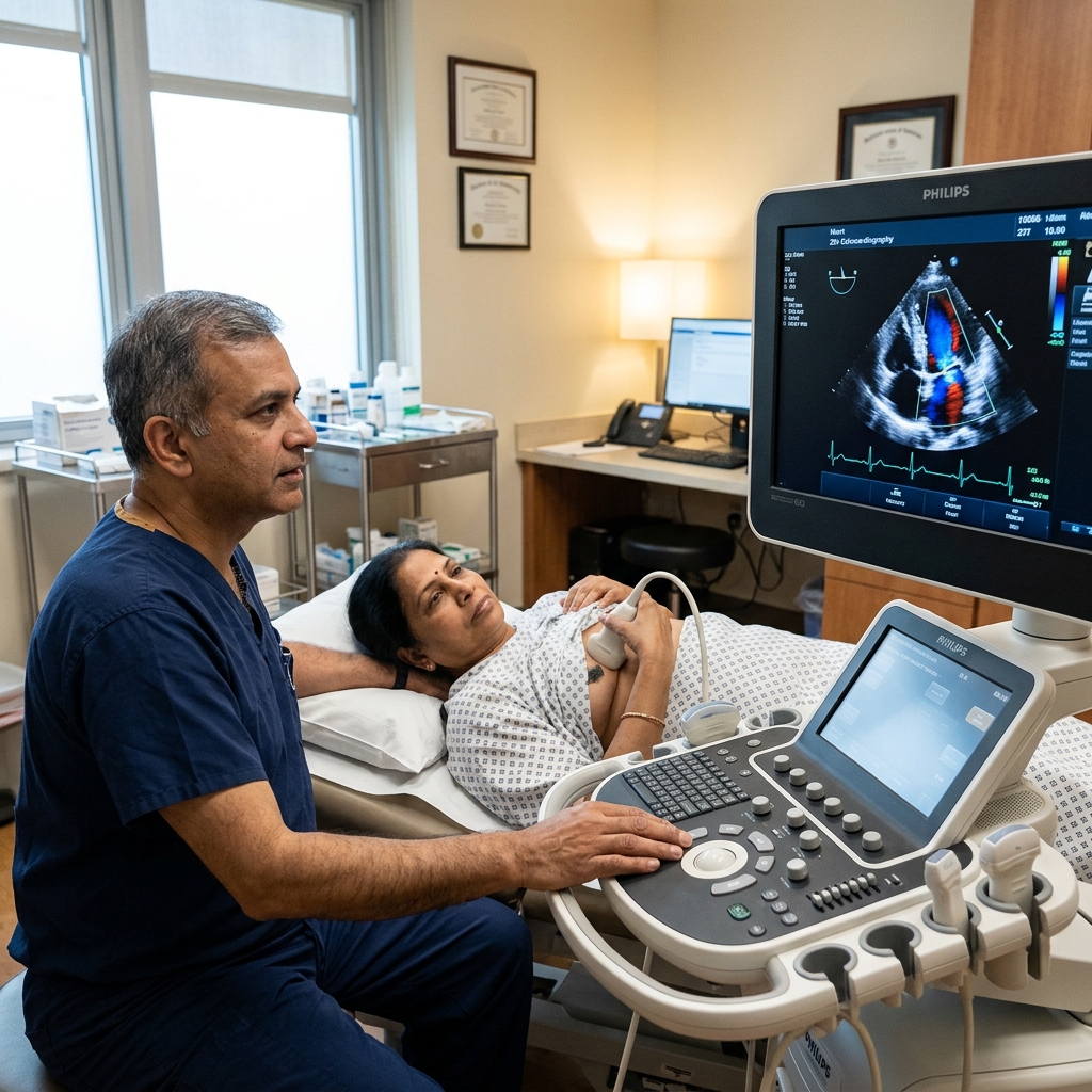
2D Echocardiography
Ultrasound imaging of heart structure, valves, and function. Complements TMT by assessing structural heart disease.
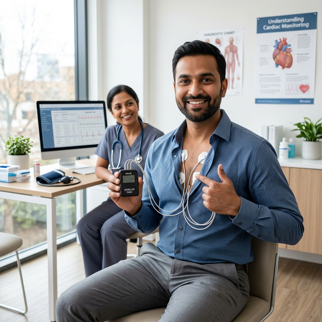
24-Hour Holter Monitoring
Continuous ambulatory ECG to detect arrhythmias that may occur spontaneously rather than with exercise.
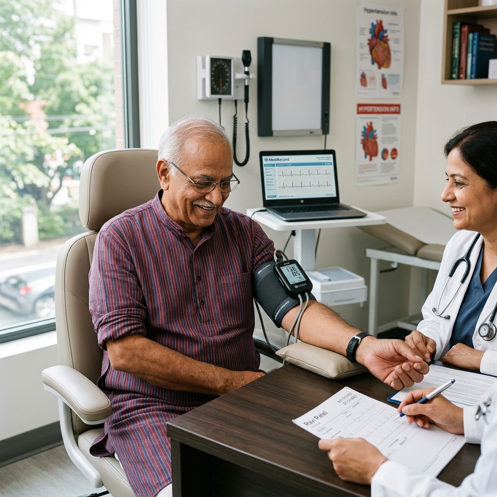
Ambulatory BP (ABPM)
24-hour blood pressure monitoring to assess hypertensive response to daily activities.

Cardiac Risk Assessment
Comprehensive 10-year cardiovascular risk profiling using SCORE2 algorithm.
Book a Visit.
Pick a date and time that works for you.
Select a date
| Su | Mo | Tu | We | Th | Fr | Sa |
|---|---|---|---|---|---|---|
Book an Appointment with Dr. Amit Singh, FACC.
Schedule a cardiology consultation at Heartwise Clinic in Vashi, Navi Mumbai — online booking, WhatsApp, or call. Dr. Amit Singh offers in-clinic and secure video teleconsultations for patients across India and internationally.
Choose Date & Time
Pick a slot that fits your schedule from available morning or evening appointments.
Share Your Details
Provide your name, contact number, and a brief note about your cardiac concern or reason for visit.
Get Confirmed
Our clinical team confirms your slot within 24 hours via call or WhatsApp with pre-visit instructions.
Consult In-Clinic or Online
Visit Heartwise Clinic in Vashi or join a secure HD video teleconsultation from anywhere.
Consultation Options
In-Clinic Consultation
Kokilaben Hospital, Kopar Khairane & Heartwise Clinic, Vashi
HD Video Teleconsultation
Available pan-India and for international patients
WhatsApp Booking
Quick booking via +91 97695 17636 — reports & follow-ups
100+ appointments this month
Confirmed by our clinical team
4.9 / 5 rating
From patient reviews
Our Cardiology
Centers.
Dr. Amit Singh consults across multiple flagship centers and outreach clinics in Navi Mumbai & Dombivli to ensure specialized, top-tier cardiac care is directly accessible.
Navi Mumbai Sectors & Surrounding Nodes Served
Triple ESC & FACC Certified
International guidelines and clinical safety protocols applied across all heart centers.
“Beat Better. Live Wiser.”
Dr. Amit Singh, FACC
Consultant Interventional Cardiologist
Medical Disclaimer: This article has been written and reviewed by Dr. Amit Singh, FACC, for educational purposes only. It does not constitute personalised medical advice and should not be used as a substitute for a consultation with a qualified cardiologist. Individual clinical decisions must be made by a treating physician based on complete medical history and examination. If you are experiencing chest pain, breathlessness, or other cardiac symptoms, seek emergency medical care immediately.