TAVR / TAVI Aortic Valve Implantation Without Surgery
A revolutionary transcatheter procedure to replace a severely calcified or narrowed aortic valve via femoral access. Discharged in 2–3 days without chest incision or cardiopulmonary bypass under Kokilaben Hospital's premier structural heart team.
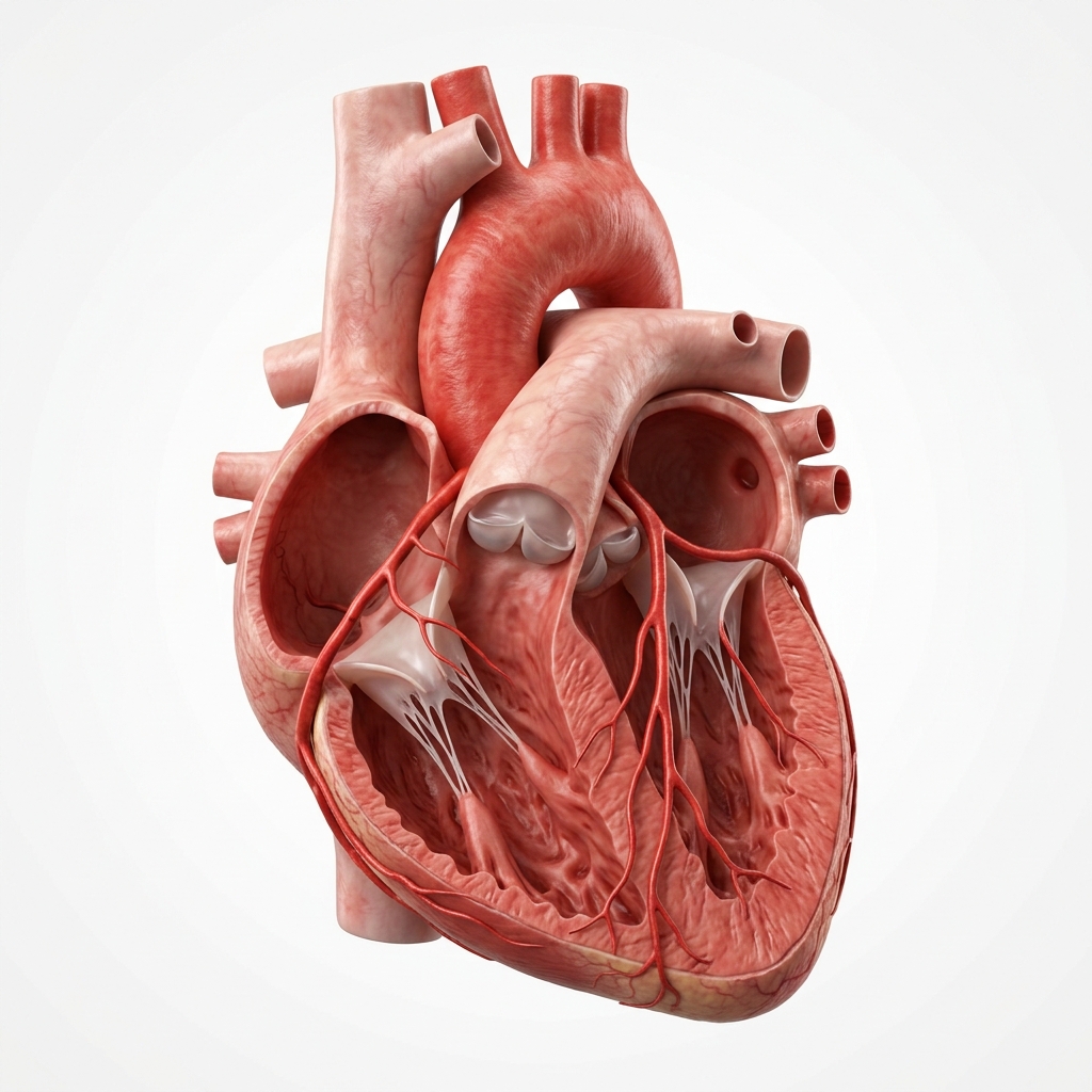
What Is Aortic Stenosis?
Understand the narrowing of the outflow tract, left ventricular loading, and the critical clinical timeline.
Aortic stenosis is a progressive disease in which the aortic valve — the outlet valve between the left ventricle and the aorta — becomes calcified and narrowed over time. As the valve area narrows, the heart must work increasingly harder to push blood through the restricted opening. Severe aortic stenosis (valve area <1.0 cm²; normal 3–4 cm²) is a life-threatening condition. Untreated symptomatic severe aortic stenosis carries a median survival of 2–3 years after symptom onset — worse than many cancers. TAVR or surgery is essential once symptoms develop.
Severe AS — Valve area <1.0 cm², Mean gradient >40 mmHg, Peak velocity >4 m/s
→ TAVR or Surgery when symptomaticModerate AS — Valve area 1.0–1.5 cm², Mean gradient 20–40 mmHg, Peak velocity 3–4 m/s
→ Annual echo surveillance; optimise risk factorsMild AS — Valve area >1.5 cm², Mean gradient <20 mmHg, Peak velocity <3 m/s
→ Monitor every 3–5 years; no interventionBalloon-Expandable vs Self-Expanding Valves
A head-to-head comparison of balloon inflation mechanics and self-expanding nitinol frames.
Balloon-Expandable Valve
Examples: Edwards SAPIEN 3, SAPIEN 3 Ultra, SAPIEN XT
Technical Specs
- •Highly predictable deployment — accurate positioning
- •Lower pacemaker implantation rate (≈5–10%)
- •Strong radial force — useful in calcified annuli
- •Excellent durability data at 5–8 years
- •Available from 20mm to 29mm; covers most annuli
Key Trial
PARTNER 3 primary trial valve: SAPIEN 3 · Outer sealing skirt: significant PVL now <1%
Self-Expanding Valve
Examples: Medtronic Evolut R, Evolut PRO, Evolut PRO+
Technical Specs
- •Fully recapturable and repositionable (Evolut PRO)
- •Supra-annular design — larger effective orifice area
- •Excellent choice for bicuspid aortic valve anatomy
- •Wide size range: 23mm to 34mm
- •Evolut Low-Risk trial: non-inferior to surgery
Key Point
Higher pacemaker rate than BE (≈15–25%) — important patient counselling point
The Importance of CT Angiography Sizing
Ensuring optimal device selection, height placement, and femoral pathway validation.
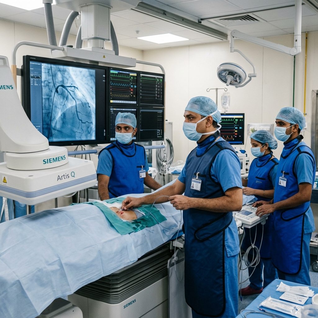
The Implantation Process in Detail
A deep clinical view into the transfemoral approach, pre-dilation pacing, and vascular closure.
Anaesthesia & Vascular Access
The transfemoral approach is preferred in >95% of cases. Two femoral access points are established: a large-bore access site (14–16Fr sheath) for valve delivery and a smaller contralateral site for haemodynamic monitoring and a temporary pacing wire. A transoesophageal echocardiography (TOE) probe is placed if general anaesthesia is used.
Aortic Valve Crossing & Pre-Dilation
A guidewire is advanced retrogradely across the diseased aortic valve into the left ventricle. Pre-dilation of the stenotic valve with a balloon catheter (balloon aortic valvuloplasty) during rapid right ventricular pacing (150–200 bpm) temporarily reduces cardiac output and stabilises the deployment zone. Some centres now perform direct TAVR without pre-dilation for selected cases.
Valve Delivery & Positioning
The compressed TAVR valve — mounted on its delivery catheter — is advanced over the guidewire from the femoral artery, up the aorta, and positioned at the native aortic annulus. Positioning is confirmed using fluoroscopy and, if available, 3D CT-fluoroscopy overlay or echocardiographic guidance. Coaxial alignment is essential.
Valve Deployment
For balloon-expandable valves: the balloon is inflated under rapid pacing (to reduce cardiac motion and output), expanding the valve frame in seconds. For self-expanding valves: the outer sheath is retracted to release the valve, which expands gradually — the system can be recaptured and repositioned if needed before full release. The new valve immediately begins functioning.
Post-Deployment Assessment
Aortography and echocardiography confirm: valve position, paravalvular leak (PVL) grade, coronary flow, mean gradient across the new valve, and any wall motion changes. If significant PVL is detected, post-dilation with a larger balloon is performed. Final haemodynamics are recorded.
Vascular Closure & Recovery
Large-bore femoral access sites are closed with percutaneous closure devices (ProGlide, MANTA). The patient is moved to the Cardiac ICU or HDU for 12–24 hours of monitoring, then transferred to the ward. Discharge is typically on day 2 or 3. ECG monitoring detects any conduction disturbance (pacemaker requirement) before discharge.
The Definitive Randomised Clinical Trials
Reviewing the historical progression from extreme high-risk cohorts to the low-risk PARTNER 3 population.
TAVR vs standard medical therapy in patients with severe AS deemed inoperable for surgery — the landmark trial that proved TAVR can save lives where surgery is impossible.
Absolute reduction in 1-year all-cause mortality: TAVR 30.7% vs standard therapy 50.7%TAVR (SAPIEN XT) vs surgery in intermediate surgical risk severe AS — established TAVR non-inferiority and opened the procedure to the much larger intermediate-risk population.
2-year death/stroke: 19.3% TAVR vs 21.1% surgery. TAVR non-inferior.TAVR (SAPIEN 3) vs surgery in low surgical risk severe AS — the definitive trial establishing TAVR as superior at 30 days and non-inferior at 1 year in the lowest-risk patients.
30-day death/stroke/rehospitalisation: TAVR vs surgery (p<0.001)Self-expanding Evolut R/PRO vs surgery in low surgical risk severe AS — confirming that TAVR's low-risk indication applies to both valve platforms, not just balloon-expandable.
24-month death/disabling stroke: 5.3% Evolut vs 6.7% surgery. Non-inferiority met.TAVR Benefits & Risks Overview
Comparing cardiac outcomes, recovery pathways, and late complications head-to-head.
Core Clinical Benefits
- Bypasses chest splitting (sternotomy) or surgical trauma
- No bypass machine or cardiac standstill necessary
- Typically discharged in 2 to 3 days
- Rapid physical recovery in 1 to 2 weeks
- Proven safe and superior across all surgical risk groups
- Significant, rapid improvements in breathlessness & energy
Potential Risks & Incidence
- Pacemaker dependence: 5-10% balloon; 15-25% self-expanding
- Paravalvular leakage (PVL): Mild common; moderate/severe <2%
- Access site hematoma or vascular complication: 2-5%
- Stroke risk: 30-day stroke approximately 2-3%
- Coronary obstruction: rare but severe (<0.5%)
Frequently Asked Questions
Detailed, peer-reviewed answers to the most common patient concerns regarding stenting and long-term care.
TAVR (Transcatheter Aortic Valve Replacement) — also called TAVI — replaces a severely narrowed aortic valve with a new bioprosthetic valve delivered through a catheter in the femoral artery in the groin, without opening the chest or stopping the heart. The new valve is compressed onto a catheter, positioned within the diseased native valve, and expanded — immediately restoring normal valve function. Hospital stay is 2–3 days. The PARTNER 3 trial (NEJM 2019) established TAVR as superior at 30 days versus surgical valve replacement even in low surgical risk patients.
TAVR is indicated for patients with severe symptomatic aortic stenosis (valve area <1.0 cm², mean gradient >40 mmHg, or peak velocity >4 m/s) with symptoms of breathlessness, chest pain, or fainting. Current 2022 ESC/EACTS and 2021 ACC/AHA guidelines recommend TAVR for all symptomatic patients over 75 (Class I recommendation), and a Heart Team decision for patients under 75. TAVR is also the preferred approach for patients at high or prohibitive surgical risk at any age.
Open-heart aortic valve surgery (SAVR) requires a sternotomy (chest opening), cardiopulmonary bypass, general anaesthesia, 7–10 days in hospital, and 6–8 weeks recovery. TAVR uses a catheter in the femoral artery — no chest opening, no bypass machine. Hospital stay is 2–3 days; most patients return to normal activity within 1–2 weeks. The PARTNER 3 trial demonstrated that TAVR produces superior 30-day outcomes vs surgery (MACE 1.0% vs 3.3%) even in low surgical risk patients, with equivalent 2-year outcomes.
Modern TAVR bioprosthetic valves have demonstrated durable function in registry data up to 10 years, with structural valve deterioration (gradual calcification of the tissue leaflets) in approximately 10–15% of patients at 10 years. If a TAVR valve does deteriorate, a second valve can be delivered inside the first via valve-in-valve TAVR — avoiding surgery entirely. Long-term durability beyond 15 years is still being established through ongoing registries. This durability question is why younger patients (<65 years) may still be offered surgical valve replacement as first choice in some cases.
TAVR is performed under sedation or general anaesthesia — patients are comfortable throughout and feel no pain during the procedure. Mild groin discomfort at the access site is common for 2–4 days after. Since there is no sternotomy (chest opening), there is no chest wound pain — a major advantage over surgery. Many patients are surprised by how little discomfort they experience. The dramatic improvement in breathing and energy levels that follows aortic valve replacement is often noticeable within days of the procedure.
After TAVR, most patients receive dual antiplatelet therapy (aspirin 75 mg + clopidogrel 75 mg daily) for 3–6 months, followed by aspirin alone lifelong. Unlike mechanical heart valves, TAVR bioprosthetic valves do not require lifelong warfarin anticoagulation unless the patient has another indication such as atrial fibrillation. For patients in atrial fibrillation, a DOAC (direct oral anticoagulant) such as rivaroxaban or apixaban may replace the antiplatelet regimen — this decision is made individually after the procedure.
TAVR Indications & Patient Selection
Current ESC/EACTS and ACC/AHA guideline recommendations for transcatheter aortic valve replacement.
TAVR vs Surgical AVR
Head-to-head comparison of transcatheter versus surgical aortic valve replacement.
Recovery After TAVR
Expected recovery trajectory after transcatheter aortic valve replacement.
In Hospital (Days 1–3)
- ICU monitoring for first 12–24 hours with continuous ECG and haemodynamic monitoring
- Bed rest for 4–6 hours after sheath removal; mobilise gradually with physiotherapy
- Echocardiogram within 24 hours to assess valve function, gradient, and paravalvular leak
- Pacemaker observation: if new LBBB or conduction abnormality, monitor for 48–72 hours
- Discharge typically on day 2 or 3 after confirming stable device parameters and mobility
First Month at Home
- No heavy lifting (>5 kg) for 2 weeks; avoid strenuous activity for 4 weeks
- Wound care: keep groin/access site dry for 48 hours; report any swelling, pain, or discharge
- Antiplatelet therapy: aspirin + clopidogrel for 3–6 months post-TAVR (per local protocol)
- Notify cardiologist if chest pain, breathlessness, palpitations, or fever develop
Long-Term Follow-Up
- Clinical review and echocardiogram at 1 month, 6 months, and annually
- Lifelong aspirin (or anticoagulation if AF develops post-TAVR)
- Endocarditis prophylaxis for dental procedures in first 6 months
- Monitor for valvular degeneration: annual echo beyond year 5
Post-TAVR Results Interpretation
Key haemodynamic and echocardiographic parameters for assessing TAVR success.
“Precision in structural interventions. Excellence in clinical outcomes.”

Dr. Amit Singh, FACC
Consultant Interventional Cardiologist
Cardiac Services Available at Heartwise
Dr. Amit Singh offers comprehensive cardiac diagnostics and management at Heartwise Cardiology Clinic, Vashi.
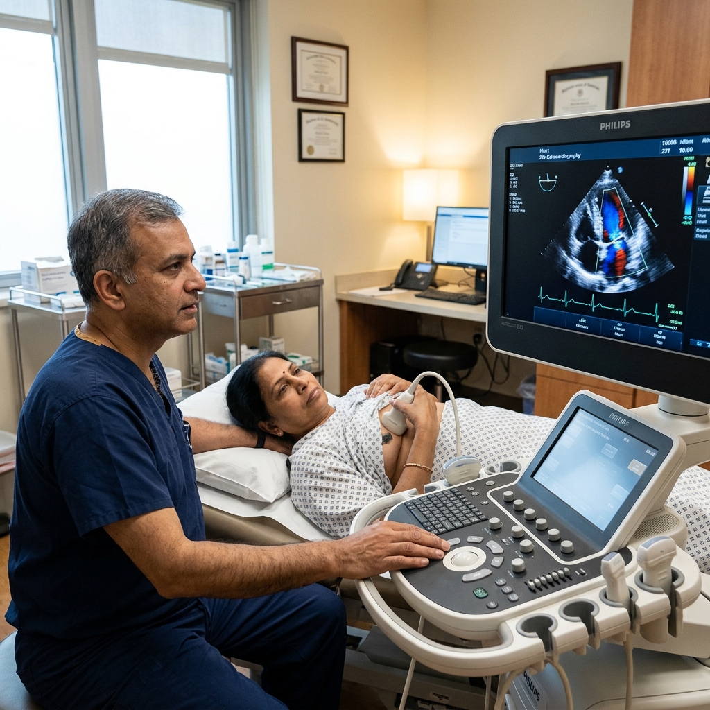
2D Echocardiography
Detailed echo assessment of AVA, mean gradient, and LV function.
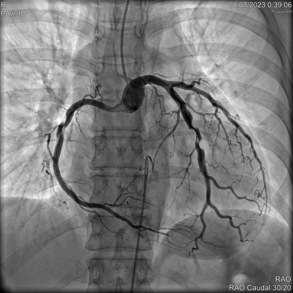
Coronary Angiography
Pre-TAVR coronary assessment — CAD frequently coexists with AS.
Aortic Stenosis
Aortic stenosis diagnosis, grading, and treatment decision-making.
Heart Failure
Post-TAVR heart failure monitoring and long-term care.
Book a Visit.
Pick a date and time that works for you.
Select a date
| Su | Mo | Tu | We | Th | Fr | Sa |
|---|---|---|---|---|---|---|
Book an Appointment with Dr. Amit Singh, FACC.
Schedule a cardiology consultation at Heartwise Clinic in Vashi, Navi Mumbai — online booking, WhatsApp, or call. Dr. Amit Singh offers in-clinic and secure video teleconsultations for patients across India and internationally.
Choose Date & Time
Pick a slot that fits your schedule from available morning or evening appointments.
Share Your Details
Provide your name, contact number, and a brief note about your cardiac concern or reason for visit.
Get Confirmed
Our clinical team confirms your slot within 24 hours via call or WhatsApp with pre-visit instructions.
Consult In-Clinic or Online
Visit Heartwise Clinic in Vashi or join a secure HD video teleconsultation from anywhere.
Consultation Options
In-Clinic Consultation
Kokilaben Hospital, Kopar Khairane & Heartwise Clinic, Vashi
HD Video Teleconsultation
Available pan-India and for international patients
WhatsApp Booking
Quick booking via +91 97695 17636 — reports & follow-ups
100+ appointments this month
Confirmed by our clinical team
4.9 / 5 rating
From patient reviews
Our Cardiology
Centers.
Dr. Amit Singh consults across multiple flagship centers and outreach clinics in Navi Mumbai & Dombivli to ensure specialized, top-tier cardiac care is directly accessible.
Navi Mumbai Sectors & Surrounding Nodes Served
Triple ESC & FACC Certified
International guidelines and clinical safety protocols applied across all heart centers.
“Beat Better. Live Wiser.”
Dr. Amit Singh, FACC
Consultant Interventional Cardiologist
Medical Disclaimer: This article has been written and reviewed by Dr. Amit Singh, FACC, for educational purposes only. It does not constitute personalised medical advice and should not be used as a substitute for a consultation with a qualified cardiologist. Individual clinical decisions must be made by a treating physician based on complete medical history and examination. If you are experiencing chest pain, breathlessness, or other cardiac symptoms, seek emergency medical care immediately.