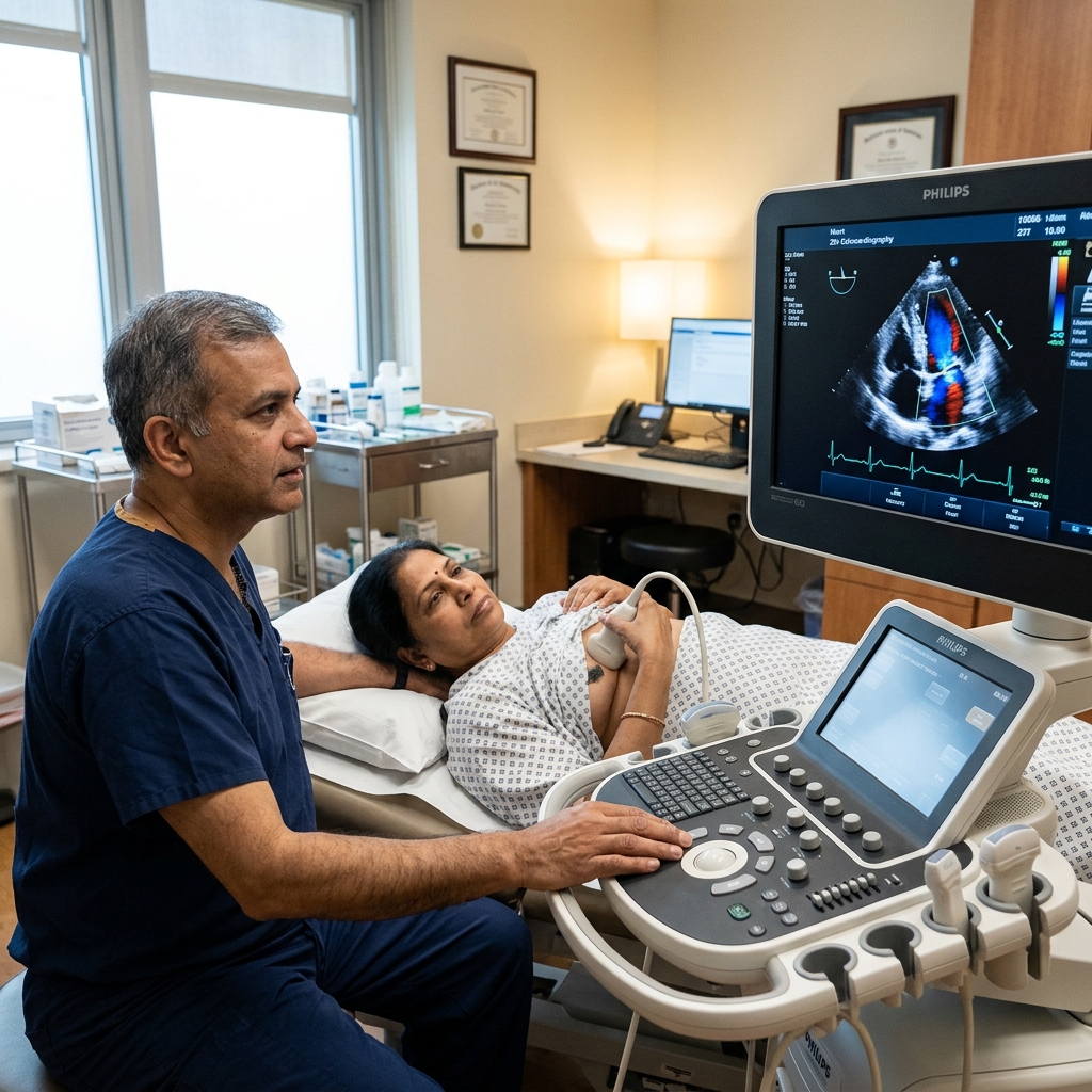
2D Echocardiography with Colour Doppler
A 2D echocardiogram uses high-frequency sound waves to produce real-time moving images of your heart — showing the four chambers, four valves, pumping muscle, and surrounding pericardium. Reported by Dr. Amit Singh, FACC.
What Does an Echocardiogram Show?
A 2D echocardiogram with Colour Doppler provides a comprehensive real-time assessment of the heart's structural and functional status.
The primary measurement is ejection fraction (EF) — the percentage of blood pumped out of the left ventricle with each beat — which determines heart pump function. Colour Doppler adds velocity and direction of blood flow through each valve, detecting leakage (regurgitation) and narrowing (stenosis) with quantitative precision. The test takes 20–30 minutes and is completely painless, using only ultrasound waves with no radiation exposure.
Causes of Cardiac Abnormalities Detected by Echo
Echocardiography reveals structural and functional changes caused by various cardiac conditions.
Coronary Artery Disease
Narrowing of coronary arteries leads to wall motion abnormalities visible on echo. Prior heart attacks produce scar tissue that appears as akinetic or dyskinetic segments.
Hypertension
Long-standing high blood pressure causes left ventricular hypertrophy (LVH), concentric remodelling, and diastolic dysfunction — all readily quantified on echocardiography.
Valvular Heart Disease
Rheumatic heart disease, degenerative calcific stenosis, and myxomatous degeneration cause valve thickening, restricted motion, or prolapse, producing regurgitation or stenosis.
Cardiomyopathy
Dilated, hypertrophic, and restrictive cardiomyopathies produce characteristic echo findings — from dilated thin-walled chambers to asymmetrical septal hypertrophy.
Pericardial Disease
Pericardial effusion, constrictive pericarditis, and pericardial thickening are well-visualised on echo. Tamponade physiology can be identified by chamber collapse.
Congenital Heart Disease
Atrial and ventricular septal defects, patent ductus arteriosus, and other congenital anomalies are identified and quantified on echocardiography from childhood through adulthood.
6 Specialized Ultrasound Modes
A comprehensive echocardiogram combines multiple ultrasound modalities, each providing a different piece of diagnostic information.
Clinical Indications: Who Needs an Echo?
Echocardiography is indicated for a wide range of symptoms, clinical conditions, and cardiac monitoring protocols.
Breathlessness & Chest Pain
Echo determines if the heart is the cause — assessing EF, diastolic function, valve disease, and pulmonary hypertension in patients presenting with dyspnoea.
Heart Murmur
Identifies which valve is affected, quantifies severity of regurgitation or stenosis, and determines whether surgical intervention is needed.
Heart Failure Suspected
Confirms diagnosis of heart failure, classifies EF (HFrEF, HFmrEF, HFpEF), and guides evidence-based treatment selection.
Post-Heart Attack (Post-MI)
Assesses EF, wall motion abnormalities, and mechanical complications such as VSD, mitral regurgitation, and pericardial effusion after myocardial infarction.
Atrial Fibrillation
Evaluates left atrial size, screens for structural causes, and assesses diastolic function in patients with atrial fibrillation.
Pre-Operative Clearance
Risk stratification before non-cardiac surgery — EF, valve function, and wall motion are assessed to guide perioperative management.
Appointment Preparation & Report Interpretation
A guide on how to prepare for the test and understand the complex numbers in your cardiology report.
Eat and Drink Normally
No fasting required. Continue all medications as prescribed before the echocardiogram.
Wear Comfortable Clothing
Wear a loose, comfortable top. You will need to remove your upper garment and wear a hospital gown for the test.
Positioning During the Test
You will lie on your left side on an examination couch. Occasionally you may be asked to lie on your back or hold your breath briefly.
What You Will Experience
A small amount of gel is applied to your chest — cool at first. The transducer is pressed gently against the chest at multiple positions. You will hear swishing sounds (Doppler signals of blood flow).
Duration and Results
The test takes 20–30 minutes. The result is discussed with you and a written report is prepared the same day.
Risks of Ignoring Echo Abnormalities
Abnormal echocardiographic findings that go unaddressed can lead to irreversible cardiac damage.
Progressive Heart Failure
A reduced ejection fraction (EF <40%) left untreated leads to progressive worsening of pump function, hospitalisations for decompensation, and increased mortality.
Valve Disease Deterioration
Moderate aortic stenosis or mitral regurgitation can progress to severe disease. Delayed intervention results in irreversible ventricular dysfunction and higher surgical risk.
Pulmonary Hypertension
Elevated RVSP on echo indicates pulmonary hypertension. Untreated, this leads to right heart failure, fluid retention, and reduced exercise capacity.
Cardiomyopathy Progression
Undetected or untreated cardiomyopathy — dilated, hypertrophic, or restrictive — progresses to end-stage heart failure, arrhythmias, and sudden cardiac death.
Treatment Guided by Echocardiography
Echo findings directly guide medical therapy, surgical timing, and device implantation decisions.
GDMT for Reduced EF
EF <40% triggers guideline-directed medical therapy (GDMT) — ACEi/ARNi, beta-blockers, MRAs, and SGLT2 inhibitors. Echo tracks response to therapy.
Valve Intervention Timing
Severe aortic stenosis with symptoms or EF <50% triggers valve replacement. Severe mitral regurgitation with LVEF ≤60% or LVESD ≥40 mm triggers repair or replacement.
Device Therapy Assessment
EF ≤35% with LBBB ≥150 ms warrants CRT assessment. EF ≤35% with prior MI or syncope warrants ICD evaluation for primary prevention of sudden death.
HFpEF Management
Heart failure with preserved EF (HFpEF) is diagnosed by echo (EF ≥50%, diastolic dysfunction, elevated filling pressures). Treatment focuses on decongestion and comorbidity management.
Common Heart Failure & Valve Disease Medications
Echocardiographic findings determine which medication classes are indicated for each patient.
Lifestyle for Heart Health
Healthy lifestyle choices complement medical therapy and improve echocardiographic parameters over time.
Heart-Healthy Diet
A Mediterranean diet rich in fruits, vegetables, whole grains, and omega-3 fatty acids supports myocardial function and reduces cardiovascular risk.
Regular Exercise
Moderate aerobic exercise for 150 minutes weekly improves exercise capacity and may slow LV remodelling in heart failure patients.
Weight Management
Obesity increases cardiac workload. Weight loss reduces LV mass, improves diastolic function, and lowers pulmonary pressures.
Smoking Cessation
Smoking accelerates coronary atherosclerosis and impairs myocardial function. Smoking cessation is the single most impactful lifestyle intervention for cardiac health.
Echocardiography Guideline Standards
International guidelines define chamber quantification standards and appropriate use criteria for echocardiography.
When to See a Cardiologist for an Echo
Certain symptoms and conditions warrant prompt echocardiographic evaluation by a cardiologist.
New or Worsening Breathlessness
Shortness of breath on minimal exertion or at night (orthopnoea, PND) requires urgent echo to assess for heart failure, valve disease, or pulmonary hypertension.
Chest Pain or Tightness
Chest pain — especially if exertional — with a murmur or signs of heart failure requires echocardiography to assess wall motion and valve function.
Heart Murmur on Examination
A new or changing murmur on physical examination warrants echocardiography to determine the cause and severity of the underlying valve lesion.
Known Heart Disease with Symptoms
Patients with known heart failure, valve disease, or prior heart attack who develop new or worsening symptoms need repeat echo to reassess cardiac function.
Frequently Asked Questions
Detailed, peer-reviewed answers to the most common patient concerns regarding echocardiography.
A 2D echocardiogram uses high-frequency sound waves (ultrasound) to produce real-time moving images of the heart's chambers, valves, and pumping muscle. Colour Doppler simultaneously maps blood flow through the valves. Together, these images show how well the heart pumps (ejection fraction), whether the valves are normal, whether the heart walls are moving correctly, and whether pressures are normal — without any radiation or needles. It is the most informative single cardiac test.
Ejection fraction (EF) is the percentage of blood pumped out of the left ventricle with each heartbeat. It is the primary measure of heart pumping function measured on echocardiography. A normal ejection fraction is 55–70%. EF 40–54% indicates mildly reduced function. EF 35–39% is moderately reduced. EF below 35% is severely reduced — this is the threshold for heart failure with reduced ejection fraction (HFrEF), at which point guideline-directed medical therapy and device therapy (CRT/ICD) are considered.
No — they are completely different tests. An ECG (electrocardiogram) records the electrical activity of the heart. A 2D echocardiogram uses ultrasound to produce images of the heart's physical structure and motion. Both are non-invasive and painless, but they provide entirely different information. An ECG shows heart rhythm and electrical conduction. An echo shows the anatomy, pumping function, valve structure, and blood flow. Most patients with heart symptoms have both tests.
A complete 2D echocardiogram with Colour Doppler at Heartwise Cardiology Clinic typically takes 20–30 minutes. No special preparation is needed — you can eat, drink, and take your medications as normal before the test. The result is available the same day and reviewed directly by Dr. Amit Singh, FACC.
No. Echocardiography uses ultrasound (high-frequency sound waves), not radiation. It is completely safe to repeat as often as needed — for monitoring heart failure, valve disease, or response to treatment. It is safe in pregnancy, in children, and in the elderly. There are no known health risks associated with diagnostic cardiac ultrasound at the frequencies used in standard echocardiography.
Minimal preparation is required. You can eat, drink, and take your medications as normal before the test. Wear a loose, comfortable two-piece outfit. Do not apply body lotions or powders to your chest area on the day of the test as they can interfere with ultrasound transmission. The test is performed with you lying on your left side, and a small amount of water-based gel is applied to your chest. The entire procedure takes about 20–30 minutes and you can resume normal activities immediately.
A standard resting echocardiogram cannot directly visualise the coronary arteries. However, it can detect the effects of blocked arteries by showing wall motion abnormalities — segments of the heart muscle that do not contract normally due to reduced blood supply. A stress echocardiogram (echo performed during exercise or with medication) is more sensitive for detecting significant coronary artery disease by comparing wall motion at rest and under stress. CT coronary angiography is the test that directly images the coronary arteries.
A transthoracic echocardiogram (TTE) is the standard test performed by moving an ultrasound probe on the surface of the chest. It is non-invasive and provides excellent images in most patients. A transoesophageal echocardiogram (TOE or TEE) involves passing a specialised probe down the oesophagus to obtain images from behind the heart. TOE provides higher-resolution images of certain structures — particularly the left atrial appendage, mitral valve, and aortic valve — and is used when TTE images are inadequate or specific clinical questions remain unanswered. TOE requires conscious sedation and fasting.
For stable heart failure with reduced EF (HFrEF), a follow-up echo is typically recommended 3–6 months after initiating or changing guideline-directed medical therapy to assess EF response. Once EF has stabilised, annual echo is usually sufficient unless symptoms change. If EF improves to >40%, medications should still be continued long-term as EF can decline again. For heart failure with preserved EF (HFpEF), routine follow-up echo is guided by symptom status rather than fixed intervals.
“Advanced cardiovascular care. Restoring life, rhythm, and vitality.”

Dr. Amit Singh, FACC
Consultant Interventional Cardiologist
Related Tests & Conditions
Echocardiography is often performed alongside other cardiac investigations for complete evaluation.
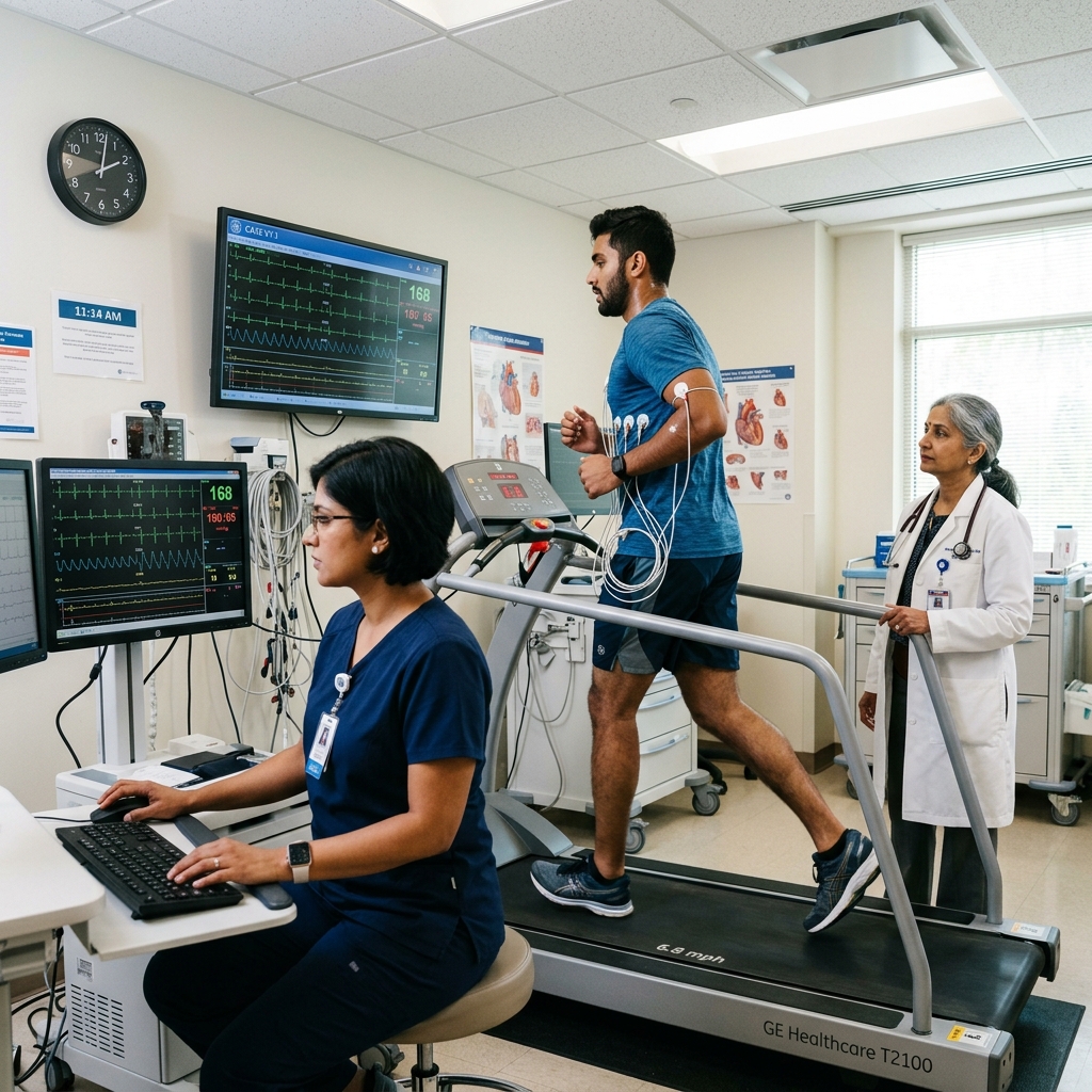
Treadmill Stress Test (TMT)
Exercise ECG to detect coronary artery disease and assess functional capacity.

24-Hour Holter Monitoring
Continuous ECG recording over 24–48 hours to detect intermittent arrhythmias and correlate with symptoms.
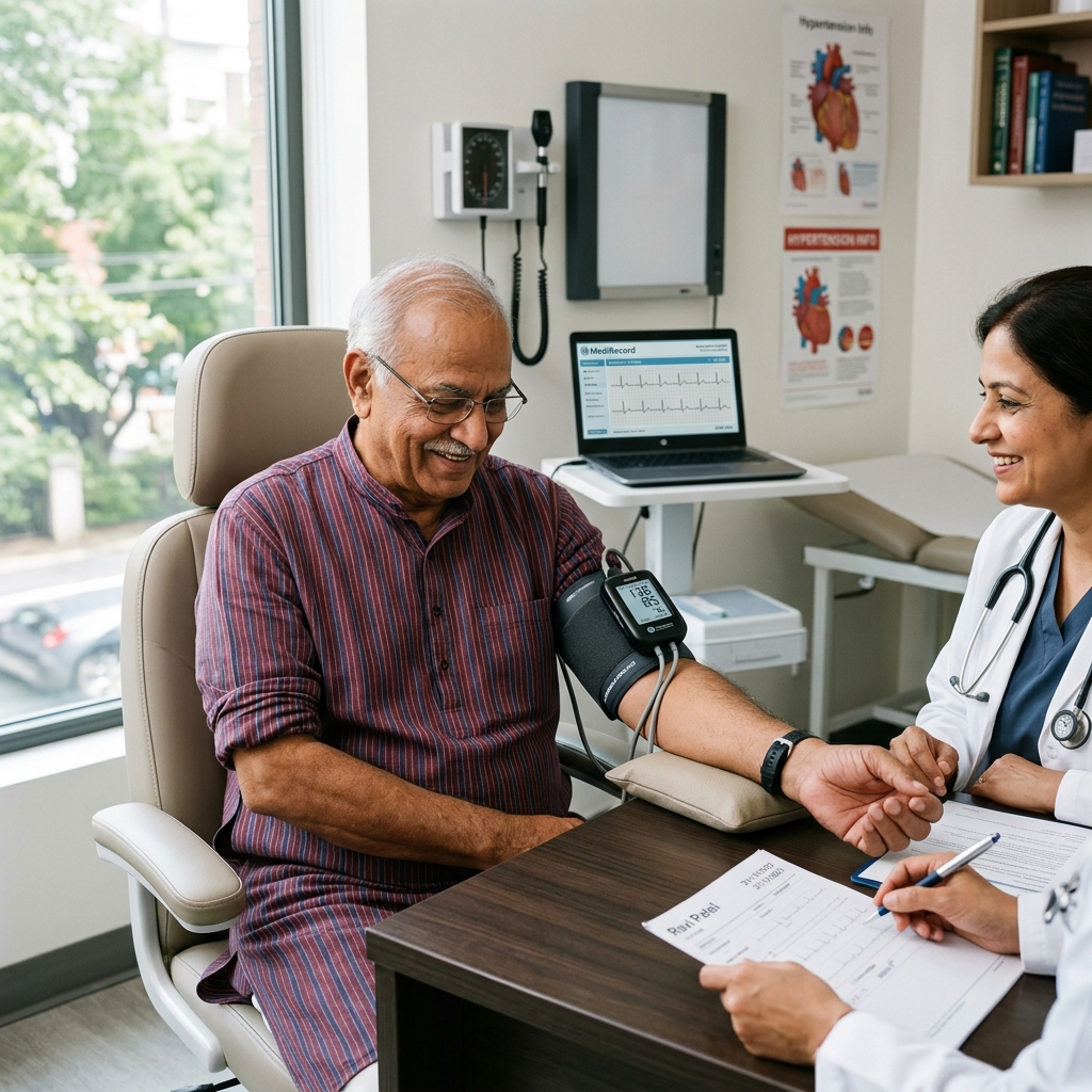
Ambulatory BP (ABPM)
24-hour automatic blood pressure monitoring that captures full diurnal BP profile.
Heart Failure
Chronic condition where the heart pumps inefficiently. Echo is essential for diagnosis and classification.
Book a Visit.
Pick a date and time that works for you.
Select a date
| Su | Mo | Tu | We | Th | Fr | Sa |
|---|---|---|---|---|---|---|
Book an Appointment with Dr. Amit Singh, FACC.
Schedule a cardiology consultation at Heartwise Clinic in Vashi, Navi Mumbai — online booking, WhatsApp, or call. Dr. Amit Singh offers in-clinic and secure video teleconsultations for patients across India and internationally.
Choose Date & Time
Pick a slot that fits your schedule from available morning or evening appointments.
Share Your Details
Provide your name, contact number, and a brief note about your cardiac concern or reason for visit.
Get Confirmed
Our clinical team confirms your slot within 24 hours via call or WhatsApp with pre-visit instructions.
Consult In-Clinic or Online
Visit Heartwise Clinic in Vashi or join a secure HD video teleconsultation from anywhere.
Consultation Options
In-Clinic Consultation
Kokilaben Hospital, Kopar Khairane & Heartwise Clinic, Vashi
HD Video Teleconsultation
Available pan-India and for international patients
WhatsApp Booking
Quick booking via +91 97695 17636 — reports & follow-ups
100+ appointments this month
Confirmed by our clinical team
4.9 / 5 rating
From patient reviews
Our Cardiology
Centers.
Dr. Amit Singh consults across multiple flagship centers and outreach clinics in Navi Mumbai & Dombivli to ensure specialized, top-tier cardiac care is directly accessible.
Navi Mumbai Sectors & Surrounding Nodes Served
Triple ESC & FACC Certified
International guidelines and clinical safety protocols applied across all heart centers.
“Beat Better. Live Wiser.”
Dr. Amit Singh, FACC
Consultant Interventional Cardiologist
Medical Disclaimer: This article has been written and reviewed by Dr. Amit Singh, FACC, for educational purposes only. It does not constitute personalised medical advice and should not be used as a substitute for a consultation with a qualified cardiologist. Individual clinical decisions must be made by a treating physician based on complete medical history and examination. If you are experiencing chest pain, breathlessness, or other cardiac symptoms, seek emergency medical care immediately.