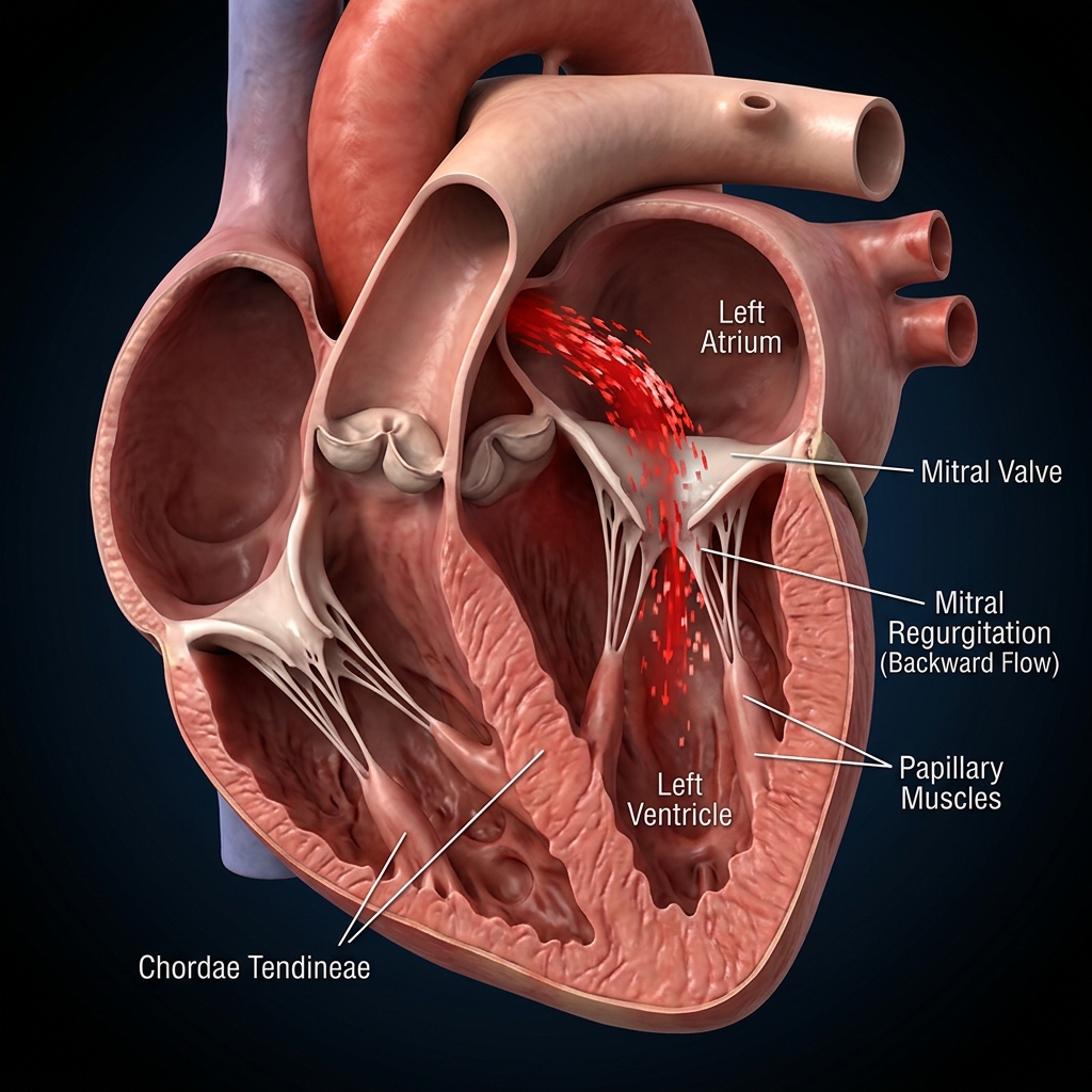
Rheumatic Heart Disease
Rheumatic heart disease develops after repeated streptococcal infections and may damage the mitral valve, aortic valve, or both, leading to valve stenosis and regurgitation.
What Is Rheumatic Heart Disease?
A progressive valve disease caused by inflammation after untreated or inadequately treated streptococcal throat infection.
Rheumatic heart disease is a chronic consequence of acute rheumatic fever. The immune response after a streptococcal infection may damage the heart valves, especially the mitral valve. Over time, this creates valve narrowing (stenosis) or leakage (regurgitation), causing breathlessness, arrhythmias, and eventual heart failure.
How It Develops
It usually follows recurrent or untreated Group A streptococcal pharyngitis in childhood.
Autoimmune Valve Injury
Molecular mimicry after streptococcal illness can trigger inflammation of the heart valves.
Mitral Valve Involvement
The mitral valve is most commonly affected, leading to stenosis and atrial enlargement.
Progressive Fibrosis
Repeated inflammation leads to fusion, calcification, and permanent structural damage over years.
Signs to Watch For
Symptoms may remain silent until the valve becomes significantly narrowed or leaking.
Breathlessness
A common early symptom that worsens with exertion and may progress to orthopnoea or pulmonary congestion.
Palpitations
Atrial fibrillation often develops as the left atrium enlarges in mitral stenosis.
Fatigue
Reduced forward flow and poor cardiac output cause fatigue and poor exercise tolerance.
How It Is Diagnosed
Echocardiography is the key diagnostic test.
Transthoracic Echocardiography
Assesses valve morphology, area, gradients, and whether regurgitation or stenosis is present.
Transesophageal Echo
Provides detailed anatomy before intervention or surgery and helps plan balloon valvotomy or repair.
Rhythm Assessment
Atrial fibrillation and pulmonary hypertension are common and require separate risk assessment.
Why Early Treatment Matters
Untreated disease can cause heart failure, AF, embolic stroke, and pulmonary hypertension.
Pulmonary Hypertension
Pressure backs up into the lungs, causing exertional breathlessness and right heart strain.
Stroke
Mitral stenosis with AF increases the risk of clot formation and embolic stroke.
Atrial Fibrillation
Left atrial enlargement makes AF common and often worsens symptoms rapidly.
Treatment Options
Treatment depends on valve lesion severity and symptoms.
Medical Therapy
Diuretics, rate control, and anticoagulation are used for symptoms and stroke prevention when AF is present.
Balloon Mitral Valvotomy
A catheter-based procedure for suitable rheumatic mitral stenosis when anatomy is favourable and the valve is not heavily calcified.
Surgical Repair or Replacement
Considered when anatomy is unsuitable for balloon treatment or when there is significant regurgitation or mixed lesion disease.
Frequently Asked Questions
Patient questions about prevention, treatment, and follow-up.
01Is rheumatic heart disease preventable?
Yes. Early treatment of streptococcal pharyngitis and prevention of recurrent rheumatic fever are key to preventing permanent valve damage.
02What is balloon mitral valvotomy?
It is a catheter-based procedure used when mitral stenosis is caused by rheumatic disease and the valve anatomy is suitable. The balloon widens the narrowed valve opening without open-heart surgery.
“Advanced cardiovascular care. Restoring life, rhythm, and vitality.”

Dr. Amit Singh, FACC
Consultant Interventional Cardiologist
Ready to Consult Dr. Amit Singh?
Schedule a clinical consultation at Heartwise Cardiology Clinic, Sector 17, Vashi or Kokilaben Hospital, Navi Mumbai.
Heartwise in Vashi. Procedures at Kokilaben.
OPD consultations, diagnostics, and patient care at Heartwise Cardiology Clinic, Vashi. Interventional procedures performed at Kokilaben Hospital, Koperkhairne.
Heartwise Clinic — Vashi
Sector 17, Vashi
Comprehensive cardiac OPD, diagnostics and consultations at the Heartwise Cardiology Clinic in Sector 17, Vashi, Navi Mumbai.
Kokilaben Hospital — Koperkhairne
Koperkhairne, Navi Mumbai
Tertiary interventional cardiac procedures — angiography, angioplasty, TAVR, structural interventions and device implantation.
Palava City — Dombivli
Palava, Dombivli
Outreach consultation clinic serving Dombivli, Kalyan and surrounding Palava City catchment.
Deepisha Medical Centre — Ulwe
Ulwe, Navi Mumbai
Cardiac OPD consultation clinic serving Ulwe, Seawoods, Belapur and the Navi Mumbai International Airport catchment.
Serving All Sectors of Navi Mumbai & MMR
Patients routinely visit our Vashi clinic from across Navi Mumbai and the surrounding Mumbai Metropolitan Region.
“Beat Better. Live Wiser.”
Dr. Amit Singh, FACC
Consultant Interventional Cardiologist
Medical Disclaimer: This article has been written and reviewed by Dr. Amit Singh, FACC, for educational purposes only. It does not constitute personalised medical advice and should not be used as a substitute for a consultation with a qualified cardiologist. Individual clinical decisions must be made by a treating physician based on complete medical history and examination. If you are experiencing chest pain, breathlessness, or other cardiac symptoms, seek emergency medical care immediately.